Painful lower extremity lesions associated with an ankle fracture
CASE
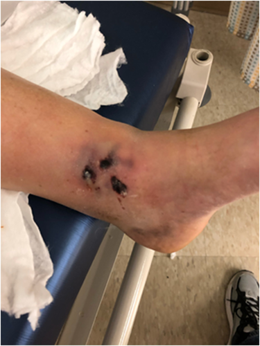
A 40-year-old female presents to day surgery for open reduction and internal fixation of the left ankle for repair of a closed trimalleolar fracture that occurred 4 days before. The ankle had initially been severely dislocated to almost 90 degrees laterally and was reduced in the field (Figure 1). There were no initial open wounds at time of the fracture. Two days after the fracture, while awaiting surgery, the patient developed severe burning pain on the medial aspect of the ankle that was unrelieved by over-the-counter and opioid medications. There were no other associated symptoms. When the dressing was removed prior to surgery, the following skin lesions were observed (Figure 2).


DIAGNOSIS
Fracture blisters
DISCUSSION
Fracture blisters are complications of fractures that look similar to second-degree burns but are actually caused by straining or shearing of the skin over the fracture site at the time of injury.1, 2 This complication occurs in approximately 3% of patients and most commonly in those with ankle, foot, elbow, and wrist injuries.1, 2 It is proposed that areas of thin, tight skin directly overlying the injury, in addition to swelling and hypoxia-induced lymphatic and venous injury, contribute to fracture blister predisposition.1, 2 Patient-specific risk factors include peripheral vascular disease, smoking, hypertension, diabetes, alcoholism, lymphatic obstruction, and collagen vascular disease.1, 2 High-energy injuries can also predispose patients to fracture blisters.1 There is not a definitive answer as to whether a fracture blister should be managed conservatively or treated with immediate versus delayed surgical repair.1-3 It is typically recommended to leave the fracture blister alone to heal completely, before proceeding with any surgery1, 3. Regardless of the treatment strategy, it is imperative to provide proper wound care and prevent bacterial infections.2 Fracture blisters tend to heal well with time; however, long-term sequelae can include scarring over the area of the blister, wound rupture, infection, and chronic ulcers.1, 2, 4


