Mitochondrial DNA: Epigenetics and environment
Abstract
Maintenance of the mitochondrial genome is essential for proper cellular function. For this purpose, mitochondrial DNA (mtDNA) needs to be faithfully replicated, transcribed, translated, and repaired in the face of constant onslaught from endogenous and environmental agents. Although only 13 polypeptides are encoded within mtDNA, the mitochondrial proteome comprises over 1500 proteins that are encoded by nuclear genes and translocated to the mitochondria for the purpose of maintaining mitochondrial function. Regulation of mtDNA and mitochondrial proteins by epigenetic changes and post-translational modifications facilitate crosstalk between the nucleus and the mitochondria and ultimately lead to the maintenance of cellular health and homeostasis. DNA methyl transferases have been identified in the mitochondria implicating that methylation occurs within this organelle; however, the extent to which mtDNA is methylated has been debated for many years. Mechanisms of demethylation within this organelle have also been postulated, but the exact mechanisms and their outcomes is still an active area of research. Mitochondrial dysfunction in the form of altered gene expression and ATP production, resulting from epigenetic changes, can lead to various conditions including aging-related neurodegenerative disorders, altered metabolism, changes in circadian rhythm, and cancer. Here, we provide an overview of the epigenetic regulation of mtDNA via methylation, long and short noncoding RNAs, and post-translational modifications of nucleoid proteins (as mitochondria lack histones). We also highlight the influence of xenobiotics such as airborne environmental pollutants, contamination from heavy metals, and therapeutic drugs on mtDNA methylation. Environ. Mol. Mutagen., 60:668–682, 2019. © 2019 Wiley Periodicals, Inc.
Abbreviations
-
- 5caC
-
- 5-carboxylcytosine
-
- 5fC
-
- 5-formylcytosine
-
- 5hmC
-
- 5-hydroxymethylcytosine
-
- 5mC
-
- 5-methylcytosine
-
- 6mA
-
- N6-methyladenine
-
- 6hmA
-
- N6-hydroxymethyladenine
-
- A
-
- adenine
-
- AGO2
-
- argonaute 2
-
- AID
-
- activation-induced deaminase
-
- ALKBH
-
- AlkB homolog
-
- APOBEC3
-
- apolipoprotein B mRNA editing enzyme catalytic polypeptide-like 3
-
- ASncmtRNA
-
- mitochondrial antisense ncRNA
-
- ATP
-
- adenosine triphosphate
-
- BER
-
- base excision repair
-
- C
-
- cytosine
-
- CHB
-
- chronic Hepatitis B virus infection
-
- CKD
-
- chronic kidney disease
-
- CO/CoX
-
- cytochrome c oxidase
-
- CYB/CYTB
-
- cytochrome B
-
- DNMT
-
- DNA methyltransferase
-
- G
-
- guanine
-
- HSP
-
- heavy strand promoter
-
- LIPCAR
-
- long Intergenic noncoding RNA predicting CARdiac remodeling
-
- LncRNA
-
- long noncoding RNA
-
- LSP
-
- light strand promoter
-
- MBD
-
- methyl-CpG-binding domain
-
- MBD4
-
- methyl-CpG-binding domain protein 4
-
- MDL1
-
- mitochondrial D-Loop 1
-
- MDL1AS
-
- mitochondrial D-Loop 1 anti Sense
-
- METTL4
-
- methyltransferase like 4
-
- miRNA
-
- microRNA
-
- mitomiR
-
- mitochondrial microRNA
-
- MMR
-
- mismatch repair
-
- mtDNA
-
- mitochondrial DNA
-
- mtSSB
-
- mitochondrial single-stranded DNA-binding protein
-
- N6AMT1
-
- N-6 adenine-specific DNA methyltransferase 1
-
- NA
-
- nucleo(t)side analog
-
- NCR
-
- noncoding control region
-
- ncRNAs
-
- noncoding RNAs
-
- ND
-
- nicotinamide adenine dinucleotide reduced dehydrogenase
-
- nDNA
-
- nuclear DNA
-
- NEIL
-
- endonuclease eight-like protein
-
- NTHL1
-
- endonuclease three-like DNA glycosylase 1
-
- NUMTs
-
- nuclear mitochondrial sequences
-
- OH
-
- origin of replication on the heavy strand
-
- OL
-
- origin of replication on the light strand
-
- OXPHOS
-
- oxidative phosphorylation
-
- PBDEs
-
- polybrominated diphenyl ethers
-
- PM
-
- particulate matter
-
- PNPase
-
- polynucleotide phosphorylase
-
- Polγ
-
- DNA polymerase gamma
-
- pre-miRNA
-
- precursor microRNA
-
- pri-miRNA
-
- primary microRNA
-
- RMRP
-
- RNA processing endoribonuclease
-
- rRNAs
-
- ribosomal RNAs
-
- SAM
-
- S-Adenosyl methionine
-
- SMUG1
-
- single-strand selective monofunctional uracil-DNA glycosylase
-
- SncmtRNA
-
- mitochondrial sense non coding RNA
-
- TDG
-
- thymidine DNA glycosylase
-
- TET
-
- ten-eleven translocation enzyme
-
- TFAM
-
- transcription factor A
-
- TFB2M
-
- mitochondrial transcription factor B2
-
- tRNAs
-
- transfer RNAs
-
- U
-
- uracil
-
- VPA
-
- valproic acid
INTRODUCTION
Mitochondrial DNA
Mitochondria are key organelles in the cell that carry out many important functions necessary for cell survival. These functions include adenosine triphosphate (ATP) production, metal homeostasis, regulation of cellular metabolism, and cellular respiration (Kaniak-Golik and Skoneczna, 2015; Lee and Han, 2017). Mitochondria are not only essential but also unique in that they contain their own DNA within the mitochondrial matrix, separate from the genomic nuclear DNA (nDNA; Yasukawa and Kang, 2018). Mitochondrial DNA (mtDNA) is circular, comprises 16,569 bp, and is double stranded where one strand is purine rich (termed the heavy strand) and the complementary strand is rich in pyrimidines (called the light strand; Asin-Cayuela and Gustafsson, 2007). First sequenced in 1981, mtDNA is inherited maternally and encodes for 13 polypeptides (subunits nicotinamide adenine dinucleotide reduced dehydrogenase [ND1-6] including ND4 and ND4L, of respiratory complex I, cytochrome c oxidase subunits I–III [CO1–3] of respiratory complex IV, subunits ATP6 and ATP8 of F1F0 ATPase, and cytochrome B [CYB] of respiratory complex III) that are required for the generation of ATP via oxidative phosphorylation (OXPHOS). mtDNA additionally encodes for 22 transfer RNAs (tRNAs) and 2 ribosomal RNAs (rRNAs; Calvo and Mootha, 2010; Yasukawa and Kang, 2018). mtDNA also possesses a noncoding control region (NCR) of ~1.1 kb that has elements to regulate replication and two major transcription initiation sites required to generate polycistronic transcripts (Shutt and Gray, 2006; Asin-Cayuela and Gustafsson, 2007). Unlike nDNA, mtDNA is not protected by arrangement into nucleosome structures; instead, it is organized into nucleoprotein complexes known as nucleoids. The primary structural constituent of mitochondrial nucleoids is mitochondrial transcription factor A (TFAM), an abundant protein, which as its name suggests, is involved with mtDNA transcription in addition to packaging mtDNA into nucleoids (Wang and Bogenhagen, 2006; Nicholls and Gustafsson, 2018). Although each mitochondrion contains multiple copies of mtDNA, the stoichiometric amounts of TFAM required for the maintenance of mtDNA has been debated (reviewed in Shokolenko and Alexeyev, 2017). However, recent super-resolution microscopy experiments suggest that each nucleoid is approximately 100 nm in diameter and comprises one copy of mtDNA packaged by multiple TFAM molecules (Kukat et al., 2011; Kukat et al., 2015).
mtDNA Replication, Transcription, and Repair
The mitochondrial genome is maintained independently of the nuclear genome by proteins that are translocated to the organelle for purposes such as DNA replication, transcription, translation, and repair. Barring the 13 polypeptides encoded by mtDNA, the mitochondrial proteome comprises an estimated 1,158 proteins as predicted by Mitocarta 2.0 (Calvo et al., 2016) that are encoded by nuclear genes and imported into the organelle via various import pathways (Becker et al., 2012; Dudek et al., 2013). The regulation of expression of these proteins could be dependent upon epigenetic, post-transcriptional, or post-translational modifications either within the nucleus or the mitochondrion.
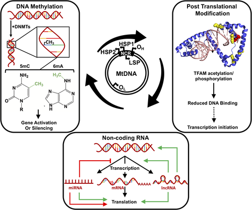
The NCR within mtDNA harbors an origin of replication on the heavy strand (OH) as well as a second origin on the light strand (OL), which is located approximately 11 kb away from OH (Fig. 1; reviewed in Young and Copeland, 2016; Falkenberg, 2018). The exact mechanism of mitochondrial replication is unknown and there are conflicting theories about the process (reviewed in Falkenberg, 2018; Yasukawa and Kang, 2018). The three main players involved with the process of mtDNA replication are DNA polymerase gamma (Polγ), the mitochondrial helicase, TWINKLE, and the mitochondrial single-stranded DNA-binding protein (mtSSB; Lee et al., 2009; reviewed in Graziewicz et al., 2006; Falkenberg, 2018; Yasukawa and Kang, 2018). The Polγ holoenzyme is a heterotrimer consisting of two identical accessory subunits (Polγ-B) and one catalytic subunit (Polγ-A), where the accessory subunits enhance the processivity and catalytic function of Polγ-A (Gray and Wong, 1992; Johnson et al., 2000; Carrodeguas et al., 2002; reviewed in Kaguni, 2004). The DNA helicase, TWINKLE, forms a hexameric donut-shaped structure and is required at the replication fork where it functions ahead of Polγ and unwinds the DNA template for replication (Korhonen et al., 2004; Milenkovic et al., 2013; Fernández-Millán et al., 2015). Until recently, Polγ was thought to be the sole polymerase within the mitochondrion that was responsible for both mtDNA replication and repair; however, at least four other DNA polymerases have been noted to play a role in mtDNA maintenance namely, DNA polymerase beta, PrimPol, polymerase theta, and polymerase zeta (Singh et al., 2015; Wisnovsky et al., 2016; Sykora et al., 2017; Bailey et al., 2019; reviewed in Bailey and Doherty, 2017; Kaufman and Van Houten, 2017; Krasich and Copeland, 2017).

mtDNA transcription is still an active area of research as several unknown variables related to initiation, elongation, and termination remain to be elucidated. As genetic information is contained within both strands of mtDNA, the bidirectionality of mtDNA transcription in Xenopus laevis was noted, a decade after the seminal work by Attardi and colleagues (Aloni and Attardi, 1971; Storrie and Attardi, 1973; Bogenhagen and Yoza, 1986). Studies involving in vitro reconstitution of mtDNA transcription minimally requires TFAM, mitochondrial transcription factor B2 (TFB2M), and mitochondrial RNA polymerase where the precise role of TFAM has been debated. Transcription of mtDNA is initiated at one of three promoter regions, two located on the heavy strand (HSP1 and HSP2) and one located on the light strand (LSP), where an overwhelming majority of transcripts are produced by transcription from HSP1 (Fig. 1; reviewed in Shokolenko and Alexeyev, 2017). Of the mitochondrial transcription components, TFAM appears to be regulated via post-translational modifications, where the precise implications of these modifications on epigenetic regulation of mitochondrial transcription are unknown (Lu et al., 2013; King et al., 2018).
Given the proximity of mtDNA to the sites of OXPHOS at the inner membrane of the mitochondria, it is not surprising that mtDNA is more prone to oxidized DNA damage when compared with its nuclear counterpart. Like the nucleus, mitochondria are also equipped with pathways to repair damaged DNA, however, not all components of nDNA repair pathways are found within the mitochondrion. For instance, the base excision repair (BER) pathway appears to be the most abundant pathway within the mitochondria and repairs small, nonbulky lesions such as alkylated and oxidized DNA bases (reviewed in Prakash and Doublié, 2015; Saki and Prakash, 2017). Only 7 of the 11 known DNA glycosylases that initiate BER have been identified in the mitochondria and recently, evidence presented by the Wilson and Bohr groups indicated that the resynthesis step after damage removal can be performed by DNA polymerase beta during repair of mtDNA (Prasad et al., 2017; Sykora et al., 2017; reviewed in Prakash and Doublié, 2015). Various proteins involved in homologous recombination and translesion DNA synthesis have also been identified within the mitochondria (Sage et al., 2010; Dmitrieva et al., 2011; García-Gómez et al., 2013; Singh et al., 2015). Although nucleotide excision repair, which is involved with the repair of bulky adducts, has not been defined as a bonafide pathway within the mitochondria, the protein Xeroderma pigmentosum group D was observed within the organelle and appears to protect mtDNA from oxidative DNA damage (Liu et al., 2015). Similarly, the proteins that initiate mismatch repair (MMR) in the nucleus have not been detected in the mitochondria, however, low levels of MMR-like activity were observed previously (Mason et al., 2003) in addition to the detection of MutL homolog 1, a protein essential for nuclear MMR (Martin et al., 2010).
Despite the presence of DNA repair pathways in the organelle, as mitochondria possess multiple copies of mtDNA, mutated mtDNA molecules exist simultaneously with undamaged mtDNA leading to heteroplasmy, blurring the line between mitochondrial disease and normalcy. Furthermore, crosstalk between the nucleus and mitochondria is essential for mitochondrial health; and because the mitochondrial proteome comprises both nuclear and mitochondrial proteins, mitochondrial dysfunction resulting from mutated nuclear proteins can also contribute to mitochondrial diseases. A growing body of evidence suggests that xenobiotic agents including environmental factors, drugs, and food additives contribute to mitochondrial disease by causing epigenetic modifications to mtDNA. In this review article, we focus our discussion on the epigenetic regulation of mtDNA as well as highlight the impact of xenobiotics on the process.
EPIGENETIC REGULATION OF mtDNA
Epigenetic changes are inheritable changes that could result in altered gene expression without any modification to the original DNA sequence. Although both nDNA as well as mtDNA can be regulated via epigenetic mechanisms, the understanding of mtDNA epigenetics has only recently gained recognition. The most common epigenetic mechanisms include noncoding RNAs and covalent modifications on either DNA or proteins. DNA can be epigenetically modified via methylation, whereas histones and other proteins can be post-translationally modified via acetylation, phosphorylation, methylation, sumoylation, ubiquitination, and PARylation. Epigenetic modifications explain differential gene expression in various cell types and tissues within an organism. These modifications can be impacted by a number of factors such as environmental exposures, which result in alterations to various cellular processes that could eventually lead to disease. Three major epigenetic mechanisms, discussed in this review article, that regulate gene expression within the mitochondrion include DNA methylation, noncoding RNAs, and post-translational modifications of nucleoid-associated proteins (Fig. 1).
The Enigma of mtDNA Methylation
DNA methylation is the addition of a methyl group from S-adenosyl-methionine (SAM) to a DNA base usually cytosine (C) or adenine (A) that is catalyzed by specialized enzymes called DNA methyltransferases (DNMTs; reviewed in Maresca et al., 2015). Although cytosine methylation occurs at position C-5 resulting in 5-methylcytosine (5mC), the exocyclic NH2 group of adenine gets methylated at position 6, which is converted into N6-methyladenine (6mA; Figs. 1 and 2). Cytosine methylation is more predominant in eukaryotes and both 5mC and 6mA in eukaryotes often result in the silencing/activation of gene transcription (Fig. 1). In the nucleus, cytosine methylation predominantly occurs within CpG dinucleotides located on CpG islands, which are repeat sequences of ~500–1500 bp in length. However, in the mitochondria, methylation occurs within CpG dinucleotides as CpG islands are virtually absent owing to the smaller size of mtDNA and its short noncoding control region. Methylation has also been observed within non-CpG (CpA, CpT, and CpC) sites within nDNA in specific cell types such as stem cells, oocytes, neurons, and glial cells (Jang et al., 2017). Adenine methylation is more common in prokaryotes and serves as a mechanism to differentiate between self and non-self DNA in order to destroy the latter. However, 6mA has also been observed in higher eukaryotes including humans (Iyer et al., 2016; Xiao et al., 2018).

Global or local DNA methylation can be detected using variations of a single or a combination of three main approaches (recently reviewed in Kurdyukov and Bullock, 2016). The first approach involves restriction digestion using methylation sensitive and insensitive enzymes followed by sequencing. The second method is affinity purification using a methyl-CpG-binding domain (MBD) or an antibody that specifically recognizes methylated bases (5mC/5hmC). The third approach uses bisulfite sequencing where the DNA is first treated with sodium bisulfite, which converts unmodified C but not 5mC to uracil (U), followed by PCR amplification and sequencing. Currently, this latter technique is considered to be the “gold standard” for DNA methylation; however, a caveat of the technique is the detection of false positives due to the incomplete conversion of unmodified cytosine to U (reviewed in Zilberman and Henikoff, 2007).
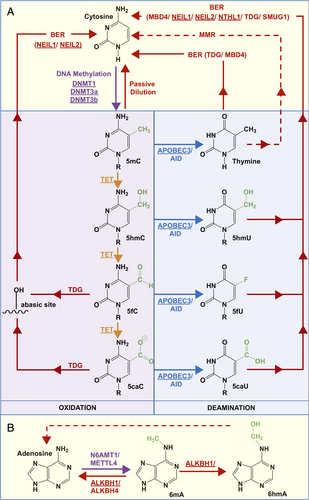
In the nucleus, the three main enzymes DNMTs, DNMT1 (methylation maintenance), and DNMT3a and 3b (de novo methylation) are responsible for cytosine methylation (Fig. 2; reviewed in Jin and Robertson, 2013; Lyko, 2018). These enzymes share a conserved catalytic motif, and genetic knockouts of DNMT1 or 3a/3b indicate that these enzymes are essential for viability (Li et al., 1992; Okano et al., 1999). Demethylation of 5mC can be achieved either by passive dilution through multiple rounds of replication or active modification (i.e., oxidation/deamination) followed by recognition by a DNA glycosylase enzyme that catalyzes the first step of DNA BER (Fig. 2A). Demethylation of 5mC is initiated via oxidation by one of the three ten-eleven translocation (TET1/2/3) dioxygenases or via deamination by activation-induced deaminase (AID) or the apolipoprotein B mRNA editing enzyme catalytic polypeptide-like 3 (APOBEC3; Fig. 2A; reviewed in Bayraktar and Kreutz, 2018). The oxidation products of 5mC by TETs form 5-hydroxymethylcytosine (5hmC), which can be further oxidized to form 5-formylcytosine (5fC) and subsequently to 5-carboxylcytosine (5caC; Fig. 2A; reviewed in Wu and Zhang, 2017; Bayraktar and Kreutz, 2018). 5fC and 5caC can then be recognized and excised by thymidine DNA glycosylase (TDG) forming an abasic (AP) site, which allows for AP endonuclease 1- or endonuclease eight-like (NEIL)-mediated BER to restore the original cytosine base (Fig. 2A; reviewed in Drohat and Coey, 2016; Bochtler et al., 2017). In one study, substrate turnover by TDG was reportedly enhanced by the NEIL enzymes (Schomacher et al., 2016). The thymine, formed due to AID or APOBEC3-mediated deamination of 5mC, can be restored to cytosine by either TDG or MBD protein 4 (MBD4)-mediated BER. 5hmC may also be deaminated to 5hmU, which can be recognized by TDG, single-strand selective monofunctional U-DNA glycosylase (SMUG1), MBD4, or the NEIL1 DNA glycosylase and repaired via BER (reviewed in Bayraktar and Kreutz, 2018). It is not clear whether active demethylation occurs via deamination of 5fC to 5fU and 5caC to 5caU, which are also known substrates of the TDG, SMUG1, endonuclease three-like DNA glycosylase 1 (NTHL1), and NEIL1 glycosylases. Enzymes associated with adenine methylation and demethylation have been recently identified in the nucleus. DNMTs, N-6 adenine-specific DNA methyltransferase 1 (N6AMT1), and methyltransferase like 4 (METTL4) can facilitate adenine methylation, whereas AlkB homolog 1 (ALKBH1) and ALKBH4 can facilitate 6mA demethylation (Fig. 2B; Xiao et al., 2018; Kweon et al., 2019). The mechanism of adenine (de)methylation remains largely unknown, and the existence of N6-hydroxymethyladenine (6hmA), an oxidation product of 6mA formed by ALKBH1, is currently acknowledged in mammalian cells and tissues (Xiong et al., 2018).
The mechanism of DNA methylation and demethylation in the mitochondria is still not clearly understood. Over the last decade, mtDNA methylation has been extensively scrutinized as several studies have identified this modification in cell lines and tissue samples of mouse and human origin, in both normal as well as disease conditions, in young versus aged mice, as well as under various conditions of oxidative stress, nutrition, and environmental exposure (Chestnut et al., 2011; Shock et al., 2011; Dzitoyeva et al., 2012; Bellizzi et al., 2013; Pirola et al., 2013; Wong et al., 2013; Ghosh et al., 2014; Baccarelli and Byun, 2015; Saini et al., 2017). Controversial reports observe either the presence or complete absence of mtDNA methylation in the noncoding control region within a short 7S DNA fragment that results in a three-stranded displacement or D-loop structure, as well as within several rRNA, tRNA, and protein encoding genes (Hong et al., 2013; Wong et al., 2013; Ghosh et al., 2014; Liu et al., 2016; Mechta et al., 2017; Mposhi et al., 2017; Matsuda et al., 2018; Owa et al., 2018). It is advantageous to measure methylation within the mitochondrial D-loop, because this region harbors the HSP and LSP elements and is easily accessible to proteins during mtDNA replication and transcription where methylation can directly impact these processes (van der Wijst et al., 2017). DNA methylation in the mitochondria was first reported almost five decades ago and has been a topic of controversy ever since (reviewed in Maresca et al., 2015; D'Aquila et al., 2017; Mposhi et al., 2017). In earlier studies in the 1970s, the presence of 5mC was reported in mouse and hamster cell lines (Nass, 1973), as well as in bovine and rat liver mtDNA (Kirnos and Vaniushin, 1976). The mtDNA of mammals, fishes, and birds displayed varying levels of 5mC in a species-dependent manner (Vanyushin and Kirnos, 1977; Nobre et al., 1978). Concurrent studies also reported the complete absence of mtDNA methylation in cultured cells and in various organisms including calf, rat, yeast, and Paramecium (Cummings et al., 1974; Groot and Kroon, 1979). Years after the first observation, in the 1980s, a study reported that 2%–5% of mtDNA at CCGG sites were fully methylated in human fibroblast cells (Shmookler Reis and Goldstein, 1983) and a separate study noted methylation in 3%–5% of CpG sites in mouse mtDNA (Pollack et al., 1984). In contrast to these observations, in 2004, one study reported the nonexistence of mtDNA methylation in cancer cell lines and tissue samples from gastrointestinal cancer patients using bisulfite-PCR–single-stranded DNA conformation polymorphism analysis and posited that mitochondrial methylation is likely a rare event (Maekawa et al., 2004). Antibodies against methylated bases 5mC and 5hmC allowed for immunoprecipitation and ELISA-like methods to detect the presence of these modifications in the mitochondria of various cultured cells and tissues (Chestnut et al., 2011; Shock et al., 2011; Chen et al., 2012; Dzitoyeva et al., 2012). In order to study the functional effect of mtDNA methylation on transcription, either bacterial CpG methyl transferase or Chorella virus GpC DNA methyl transferase were targeted to the mitochondria and while cytosine was methylated at both CpG and GpC sites, a decrease in HSP1 and HSP2 regulated gene expression was observed only in the context of GpC methylation (van der Wijst et al., 2017). In another study, overexpression of DNMT1 resulted in the modified transcription of particular genes, where MT-ND6 expression from LSP was downregulated, MT-ATP6 and MT-CO1 expression were unaltered, and expression of MT-ND1 from HSP was upregulated (Shock et al., 2011).
It has been suggested that the levels of mitochondrial methylation observed initially were overestimated due to inefficient bisulfite conversion of circular supercoiled mtDNA, and it was recommended that mtDNA should be linearized prior to the detection of methylation (Liu et al., 2016). mtDNA methylation levels may also be overestimated due to either nDNA contamination in mtDNA preparations or nuclear mitochondrial sequences (NUMTs), which are regions of the mitochondrial genome that translocated to the nuclear genome over time and thus share a high-sequence homology with their mitochondrial counterpart (Woischnik and Moraes, 2002). A recent study using bisulfite sequencing after mtDNA linearization and whole-genome shotgun sequencing did not detect significant levels of CpG and non-CpG methylation. Experimental conditions such as primer design, incomplete bisulfite conversion, and template purity/topology appear to play a critical role in the precise analysis of mtDNA methylation (Owa et al., 2018).
To date, most studies aimed at identifying methylation in mtDNA have focused efforts on determining methylation at CpG sites; however, non-CpG methylation and adenine methylation within mtDNA have also been observed (Bellizzi et al., 2013; Bianchessi et al., 2016; Blanch et al., 2016; Koh et al., 2018). The presence of 6mA in the human mitochondrial genome was reported in 2018 using 6mA-crosslinking-exonuclease-sequencing. The mitochondrial genome was found to contain >8,000 times higher 6mA than the nuclear genome, usually present within a “6mAT” dinucleotide motif. The distribution of 6mA was found to be uniform throughout mtDNA with no bias toward clusters. The functional implications of 6mA were associated with mtDNA melting and the subsequent recruitment of mtSSB to mtDNA (Koh et al., 2018). The endosymbiotic theory of mitochondrial origin combined with the presence of an ~8,000-fold increase in 6mA compared with nDNA indicates the possibility that mtDNA methylation occurs predominantly on adenine. The numerous conflicting reports surrounding cytosine methylation further supports this hypothesis.
The earliest evidence of DNMT activity in mitochondria was observed in vertebrates including loach embryos, rat liver, ox, as well as in mouse and hamster cells (Vanyushin et al., 1971; Vanyushin and Kirnos, 1976; Vanyushin and Kirnos, 1977). The expression of various DNMTs in different cell types is also controversial and appears to be preferential depending upon the cell and tissue type being studied (Saini et al., 2017; reviewed in Maresca et al., 2015). Currently, DNMT1, DNMT3a, DNMT3b, and two TET enzymes (TET1 and TET2) have been identified within the mitochondria using immunofluorescence and western blotting procedures (Chestnut et al., 2011; Shock et al., 2011; Chen et al., 2012; Dzitoyeva et al., 2012; Bellizzi et al., 2013; Wong et al., 2013; reviewed in Maresca et al., 2015). The methyl transferase responsible for adenine methylation in mitochondria has not yet been identified, however, the demethylase ALKBH1 was identified in the mitochondria in 2018 where it plays a role in regulating OXPHOS (Koh et al., 2018). The presence of TET1 and TET2 in the mitochondria indicates that mtDNA demethylation occurs via oxidation-mediated active demethylation (Fig. 2). Whether deamination-mediated demethylation takes place in mitochondria is not clear. APOBEC3 has been recently identified in the mitochondria implicating the possibility of deamination of 5mC in mtDNA (Wakae et al., 2018). The main DNA glycosylases involved with the removal of the oxidation/deamination products of 5mC include TDG as well as two other DNA glycosylases, SMUG1 and MBD4, are absent in the mitochondria (reviewed in Prakash and Doublié, 2015). Therefore, the next steps in the repair of oxidation/deamination products of methylated bases remain an area for further research and raise the possibility that other DNA glycosylases such as the NEIL1, NEIL2, or NTHL1 enzymes that are present in the mitochondria could compensate for the lack of TDG in the mitochondria and initiate the repair process. A recent study suggested that the NEIL1 enzyme can recognize and excise 5caC (Slyvka et al., 2017), a result that has been previously disputed (Fig. 2A; Moréra et al., 2012). A separate study reported that 5hmU and 5fU, the further oxidation/deamination products of 5mC, are also known substrates for NEIL1 (Fig. 2A; (Zhang et al., 2005). Yet, another possible avenue of repair for the thymine base generated opposite guanine (G) as a result of deamination of 5mC could be mismatch DNA repair-like activity that can recognize and repair G-T mismatches, but this still remains to be elucidated (Fig. 2A; Mason et al., 2003; de Souza-Pinto et al., 2009).
Impact of Xenobiotics on mtDNA Methylation
Mitochondrial dysfunction caused by alteration and damage to mtDNA is believed to be one of the major underlying mechanisms in the development of disease. With the acceptance that epigenetic modifications such as methylation play a role in mtDNA regulation, the effects of xenobiotic agents on the levels of mtDNA methylation, in particular, 5mC and 5hmC levels, have also been explored. mtDNA methylation can be affected by exposure to various external factors including air pollutants, metals, cigarette smoke, dietary oils, food supplements, and therapeutic drugs.
Airborne pollution and cigarette smoke can cause various diseases including birth defects, cardiovascular diseases, respiratory diseases, neurological disorders, and cancer (Stingone et al., 2014; Fetterman et al., 2017; Boovarahan and Kurian, 2018). Airborne particulate matter (PM) can be classified into three categories based on their size: coarse (PM10 < 10 μm diameter), fine (PM2.5 < 10 μm diameter), and ultrafine or nano (PM0.2 < 0.2 μm). These particulates can methylate the mitochondrial D-loop region and mitochondrial genes leading to an increased risk for disease. For example, the exposure of steel workers to PM10 has been associated with hypermethylation of the MT-TF (phenylalanine tRNA) and MT-RNR1 (12S rRNA) genes in peripheral blood (Byun et al., 2013). Similarly, prenatal exposure to PM2.5 in air pollution has been correlated with increased placental mtDNA methylation in the D-loop and MT-RNR1 (Janssen et al., 2015 ). In contrast, the exposure of PM2.5, produced from welding, decreases blood mtDNA methylation in the D-loop region (Byun et al., 2016). In another study, exposure to fumes from welding has been linked to hypomethylation in the D-loop and MT-TF and increases blood pressure (Xu et al., 2017). Lastly, smoking during pregnancy has been associated with methylation of the placental D-loop region as well as MT-RNR1 gene impacting health of newborns (Armstrong et al., 2016; Janssen et al., 2017).
Metal ions in the environment such as chromium and arsenic that accumulate due to occupational exposure or polluted drinking water can alter patterns of mtDNA methylation. Hypomethylation in the MT-TF and MT-RMR1 genes was observed following chromium exposure in chrome plating workers (Yang et al., 2016). Likewise, arsenic exposure was associated with hypomethylation of the D-loop and MT-ND6 in an Indian population exposed to high concentration of arsenic present in drinking water (Sanyal et al., 2018). Furthermore, arsenic exposure also resulted in an increase in mtDNA copy number, upregulation in the expression of the nuclear-encoded TFAM gene, and increased expression of the mitochondrial proteins ND4 and ND6 (Sanyal et al., 2018). Another environmental agent used in building materials, electronics, furniture, vehicles, and plastics known as polybrominated diphenyl ethers (PBDEs) are flame retardant substances that can cause neurological impairment. Upon exposure to PBDEs, a decrease in 5mC levels within the MT-CO2 gene was observed and may likely contribute to neuronal toxicity (Byun et al., 2015).
Maternal diet can alter the mtDNA methylation levels of newborns, modulating their mitochondrial OXPHOS capacity, which may have long-term consequences on energy homeostasis. For example, protein deficiency during pregnancy alters mtDNA methylation levels in a sex-specific manner and betaine supplementation has been implicated in hypomethylation in the D-loop region and overexpression of mitochondrial encoded OXPHOS genes in newborn piglets (Jia et al., 2013; Jia et al., 2015). Dietary lipid concentration can also affect mtDNA methylation as observed in large yellow croaker fish, where high lipid diet increases D-loop methylation, but low lipid diet reduces methylation in MT-RMR1 (Liao et al., 2016). Similarly, the consumption of olive oil and perilla oil increases methylation in MT-ND4L and MT-TR (arginine tRNA) in the liver of large yellow croaker. However, methylation of MT-RNR1 was decreased upon intake of perilla oil. The modulation of lipid metabolism in response to high lipid content present in these oils has been suggested to impair mitochondrial function (Liao et al., 2016).
Pharmaceutical agents can cause mitochondrial dysfunction by altering expression of DNMT/TET enzymes and consequently causing changes in the levels of mtDNA methylation. Valproic acid (VPA), an anticonvulsant and mood stabilizer, is often used in the treatment of epilepsy and may cause liver toxicity due to mitochondrial dysfunction (Silva et al., 2008). In order to understand the underlying mechanism of hepatotoxicity, the effects of VPA on mtDNA methylation were tested using mammalian cultured cells. A reduction of 5hmC levels in mtDNA was observed following VPA treatment on mouse fibroblast cells and was associated with decreased mRNA and protein levels of TET1 (Chen et al., 2012). In a separate study using primary human hepatocytes, hypomethylation of seven mitochondrial genes was observed upon VPA treatment and within the same study, two nuclear genes crucial for DNA methylation, DNMT and MAT (methionine adenosyltransferase), were found to be hypermethylated, resulting in reduced levels of the DNMT enzymes and SAM. This may impact mtDNA methylation downstream, which is suggestive of crosstalk between the nucleus and mitochondria after VPA exposure (Wolters et al., 2017). The downstream molecular events after VPA exposure were also explored in human hepatocytes, where upregulation of DNMT caused transient hypermethylation in mtDNA and downregulation of mitochondrial proteins resulting in impaired mitochondrial function. In this study, crosstalk between the nucleus and mitochondria was observed where the mitochondrial MT-CO2 gene initiates a complex cascade of interactions mediated by the nuclear genes FN1, MYC, and CPT1A. Most of these genes participate in various mitochondrial functions such as electron transport, apoptosis, mitochondrial import, and fatty acid oxidation, the impairment of which can lead to mitochondrial dysfunction (Wolters et al., 2018).
Epigenetic events are also involved in adaptive responses against chemotherapeutic drugs. A low dose of a chemotherapeutic drug, doxorubicin, downregulates DNMT1 expression in rat cardiac myocytes, resulting in mtDNA hypomethylation, which leads to upregulation of mitochondrial encoded proteins (Ferreira et al., 2019). This low dose of doxorubicin treatment increases cellular resistance against a higher subsequent dose and was believed to be an epigenetic-linked mitochondrial adaptive response facilitating effective anticancer treatment (Ferreira et al., 2019). Another therapeutic drug that elicits mitochondrial dysfunction via changes in epigenetic markers is the antiviral nucleo(t)side analog (NA), which is used to treat patients with chronic Hepatitis B virus infection (CHB). A potential cause of NA-induced mitochondrial pathogenesis may be the epigenetic changes observed in CHB patients, such as hypermethylation of the mitochondrial D-loop region that correlates with overexpression of DNMT1 (Madeddu et al., 2017).
Mitochondrial Gene Regulation by Noncoding RNAs
Noncoding RNAs (ncRNAs) are RNA molecules that lack the ability to be translated into proteins (reviewed in Mattick and Makunin, 2006). The most common ncRNAs are tRNAs and rRNAs, which are essential for protein synthesis. Other ncRNAs such as long and small ncRNAs regulate gene expression through epigenetic mechanisms and can also act as signaling molecules to regulate essential cellular and biological processes (reviewed in Huang and Zhang, 2014; Wei et al., 2017). ncRNAs involved in the epigenetic regulation of mitochondrial gene expression can be encoded by both the nuclear and mitochondrial genomes, however, it is not clear whether the mtDNA encoded ncRNAs are derived from mitochondrial genes integrated into the nuclear genome (i.e., NUMTs) or are actually transcribed inside the mitochondria (reviewed in Vendramin et al., 2017). The levels of ncRNAs can be altered manipulating cellular and mitochondrial functions during various types of cancer making them a potential target for cancer therapy (reviewed in Bienertova-Vasku et al., 2013; Matsui and Corey, 2017; Sas-Chen et al., 2017; Olavarria et al., 2018; Zhao et al., 2018).
Long Noncoding RNAs
Long noncoding RNAs (lncRNAs) encode for transcripts that are >200 bp and have a characteristic structure containing multiple domains for RNA binding, protein binding, and possibly DNA binding (reviewed in Mercer and Mattick, 2013). Similar to mRNA, they can also undergo post-transcriptional modifications such as alternative splicing, 5′ end capping, and 3′ polyadenylation (reviewed in Mercer and Mattick, 2013). Several lncRNA encoded by the mitochondrial genome have been identified and participate in the regulation of mitochondrial gene expression or act as retrograde signaling molecules to maintain activity of nuclear genes (reviewed in Dong et al., 2017; Zhao et al., 2018). The first three mtDNA encoded lncRNA identified were a mitochondrial sense ncRNA (SncmtRNA) and two antisense ncRNAs (ASncmtRNAs-1 and -2), and they contain sense and antisense strands of 16s rRNA gene, respectively, connected to a 5′ leader fragment derived from respective complementary strands. These lncRNAs are transported into the nucleus as retrograde signaling molecules and play an important role in mito-nuclear crosstalk. Both sense and antisense lncmtRNAs are overexpressed in normal proliferating cells; however, in tumor cells, SncmtRNAs are upregulated, whereas ASncmtRNAs are downregulated indicating that SncmtRNAs function in cell cycle progression while ASncmtRNAs may act as tumor suppressors (Villegas et al., 2007; Burzio et al., 2009; Landerer et al., 2011). The knockdown of ASncmtRNAs induces apoptotic cell death of cancer cells via reduced expression of an apoptosis inhibitor, surivivin, suggesting that ASncmtRNAs are promising targets for cancer therapy (Vidaurre et al., 2014). After the initial discovery, three more mtlncRNAs, ND5, ND6, and CYB were identified, which are encoded by their respective mitochondrial genes. The expression of these lncRNA is regulated by the nuclear encoded mitochondrial ribonuclease P complex. The exact function of these lncRNAs is not known, however, they form intermolecular duplexes with their functional mRNA counterparts, indicating their role in stabilizing mRNA or regulating their expression (Rackham et al., 2011). In another study, an attempt was made to identify the potential role of lncRNA as biomarkers in heart disease. Transcriptome analysis revealed the abundance of seven putative lncRNAs encoded by the mitochondrial genome, out of which, two lncRNAs were found to be significantly downregulated in heart patients. One studied of these two lncRNAs, called LIPCAR (Long Intergenic noncoding RNA predicting CARdiac remodeling; Kumarswamy et al., 2014), is a chimeric lncRNA molecule encoded by the antisense regions of the MT-CYB and MT-CO2 genes (reviewed in Dong et al., 2017). LIPCAR levels were increased in patients with late-stage myocardial infarction, suggesting that it may be a putative biomarker for chronic cardiovascular diseases and heart failure mortality (Kumarswamy et al., 2014). Two additional mtlncRNA, MDL1 (Mitochondrial D-Loop 1) and MDL1AS (MDL1 Anti Sense), were identified recently where MDL1 covers the antisense region of the tRNAPro gene and the entire mitochondrial D-loop region, whereas MDL1AS is the antisense transcript of MDL1. These lncRNAs were proposed to be precursors of transcription initiation RNAs and are thought to play a role in the regulation of mitochondrial gene expression; however, their exact function is yet to be determined (Gao et al., 2018).
Some lncRNA molecules are also encoded by nDNA and transported into the mitochondria where they can interact with mitochondrial proteins and regulate mitochondrial biosynthesis, metabolism, and apoptosis. One such lncRNA is the RNA component of the RNA processing endoribonuclease (RMRP) that is transported by RNA-binding proteins human antigen R, G-rich RNA sequence binding factor 1, and polynucleotide phosphorylase (PNPase) in the mitochondria, where it participates in mtDNA replication and transcription. Lowering the RMRP levels led to a reduction in the basal oxygen consumption rate as well as levels of the ATP6, COX1, and CYTB proteins (Wang et al., 2010; Noh et al., 2016).
Small Noncoding RNAs
Most common small noncoding RNAs studied within the mitochondria are microRNAs (miRNAs). miRNAs are usually transcribed in the nucleus from the noncoding DNA regions as primary miRNA transcripts (pri-miRNA), which is then processed by Drosha/DGC8 complex to form precursor miRNA (pre-miRNA). The pre-miRNAs are then transported into the cytoplasm by exportin 5 and further processed by DICER to form mature miRNAs in the cytoplasm. The mature miRNAs then associate with argonaute 2 (AGO2) and localize to their respective target mRNA sites (reviewed in Macfarlane and Murphy, 2010). They bind to the 3′- untranslated region of the target mRNA via sequence complementarity and recruit the RNA-induced silencing complex that either represses translation or promotes the degradation of the target mRNA resulting in decreased protein expression (reviewed in Macfarlane and Murphy, 2010). In some cases, additional mechanisms of function are also proposed where miRNA can activate transcription or upregulate protein expression (discussed in Ramchandran and Chaluvally-Raghavan, 2017; Vaschetto, 2018). The miRNAs identified in the mitochondria are collectively termed mitochondrial microRNAs (mitomiRs; Bandiera et al., 2011). mitomiRs are short single-stranded RNA molecules ~17–25 bp nucleotides in length that are encoded by the nuclear genome and translocated into the mitochondria, however, a small subset can also be encoded by the mitochondrial genome (Sripada et al., 2012; Ro et al., 2013). The exact mechanism of nuclear encoded mitomiRs transport is largely unknown, and some studies suggest that PNPase or AGO2 may be involved with the transport process (Wang et al., 2010; Bandiera et al., 2011; Zhang et al., 2014; Shepherd et al., 2017). mitomiRs regulate expression of proteins encoded by nuclear as well as mtDNA and for the purposes of this review, we will discuss only the latter (reviewed in Bandiera et al., 2013; Srinivasan and Das, 2015). mitomiRs can enhance or repress protein expression at both transcriptional and translational levels resulting in the modulation of mitochondrial metabolic activity and cellular homeostasis. For instance, specific base pairing between miR-2392 and mtDNA in an AGO2-dependent manner inhibits mtDNA transcription partially, resulting in the downregulation of OXPHOS and the concomitant upregulation of glycolysis. This metabolic reprogramming is believed to trigger chemo-resistance during cisplatin chemotherapy in tongue squamous cell carcinoma (Fan et al., 2019). At the translational level, mitomiRs downregulate most mtDNA encoded proteins. For example, miR-181c downregulates MT-CO1 by targeting the 3′ end of its mRNA resulting in the remodeling of complex IV and leading to cardiac dysfunction (Das et al., 2012; Das et al., 2014). Similarly, miR-378 overexpression in Type I diabetes downregulates the mitochondrial encoded F0 component MT-ATP6 (Jagannathan et al., 2015). Another mitomiR, miR-214, has been implicated in the pathogenesis of chronic kidney disease (CKD) by disrupting OXPHOS due to downregulation of MT-ND4L and MT-ND6 and is suggested to be a potential therapeutic target and biomarker for CKD (Bai et al., 2019).
Some mitomiRs can also upregulate the expression of mitochondrial genes; for instance, the upregulation of MT-CO1 and MT-ND1 was observed by the translocation of miR-1 and miR-1a-3p to the mitochondria (Zhang et al., 2014; He et al., 2019). Another case of enhanced translation by a mitomiR was observed in spontaneous hypertensive rats, where, upon reduced levels of the CYTB protein, miR-21 is overexpressed and translocated inside the mitochondria to counteract the downregulated CYTB protein level. Exogenous expression of miR-21 also increased the CYTB protein level without changing the mRNA levels. This was believed to be a compensatory mechanism against high blood pressure and could be used in the treatment of hypertension-associated diseases (Li et al., 2016).
Post-Translational Modifications of Mitochondrial Nucleoid Proteins
In the nucleus, epigenetic changes in the form of post-translational modifications of histone proteins alter gene transcription and maintain cell-type-specific expression patterns. These modifications include lysine methylation, phosphorylation, acetylation, ubiquitylation, sumoylation, and PARylation (reviewed in Bannister and Kouzarides, 2011). In the mitochondria where histone proteins are absent, epigenetic changes to nucleoid proteins play an important role in the regulation of mtDNA gene expression. It is estimated that approximately 63% of proteins that are localized within the mitochondrion contain lysine acetylation sites, and in one study conducted in 2011, 216 phospho-peptides were identified from mitochondrial preparations (Zhao et al., 2011). This latter number is likely higher given the increasing number of proteins that have been identified within the mitochondrion as the study was conducted. Mitochondrial proteins can be acetylated both enzymatically and nonenzymatically. Nonenzymatic acetylation is favored due to a high concentration of acetyl CoA and alkaline pH in the mitochondria. The enzymatic acetylation inside mitochondria has been recently acknowledged as four acetyl transferases ACAT1, MOF, GCN5L1, and PCAF have been identified in the mitochondria and are responsible for regulation of acetylation levels of mitochondrial proteins (Fan et al., 2014; Chatterjee et al., 2016; Wang et al., 2017; Savoia et al., 2019). Deacetylation is predominantly performed by the NAD+-dependent sirtuin family of enzymes, specifically SIRT3, whereas two-additional mitochondrial sirtuin enzymes, SIRT4 and SIRT5, remove longer acyl groups including succinyl, malonyl, and lipoyl groups (reviewed in He et al., 2012). A number of kinases and protein phosphatases have also been identified within the mitochondria and regulate protein phosphorylation and dephosphorylation (reviewed in Lim et al., 2016).
TFAM, the main structural constituent of mitochondrial nucleoids is one example of a mitochondrial protein that gets post-translationally modified via acetylation, O-linked glycosylation, and phosphorylation (Suarez et al., 2008; Lu et al., 2013; King et al., 2018). Two serine residues (S55 and S56) within the high-mobility group 1 (HMG1) domain are primary sites for phosphorylation, whereas four lysine residues (K62, K76, K111, and K118) also within HMG1 appear to be sites for acetylation (Fig. 1). Phosphorylation and acetylation both regulate aspects of mtDNA dynamics by altering the binding affinity of TFAM to DNA where reduced binding of TFAM to DNA causes increased diffusion of the protein on DNA and decreased mtDNA compaction. The extent to which mtDNA is compacted ultimately influences mtDNA replication and transcription. Acetylation and phosphorylation sites have also been identified in other mitochondrial nucleoid-associated proteins including mtSSB and DNA Polγ however, the exact role of these post-translational modifications in regulating mtDNA metabolism has not been elucidated thus far (Matsuoka et al., 2007; Choudhary et al., 2009; Zhou et al., 2013).
CONCLUDING REMARKS
Mitochondria are perhaps one of the most intriguing organelles within a cell. Not only do they contain their own DNA that is maternally inherited but their endosymbiotic origin makes them evolutionarily unique (reviewed in Gray, 2012). The double-membraned organelle, much like its prokaryotic ancestor, is capable of supporting processes such as replication, protein production to generate ATP, and possesses the machinery to drive its own division. Although only a small fraction of proteins found within the organelle are encoded by mtDNA, a majority of the mitochondrial proteome is encoded within the nucleus making crosstalk between the two organelles necessary and inevitable. Furthermore, depending upon the cell type, many mitochondria exist within a single cell and each mitochondrion has multiple copies of mtDNA leading to heteroplasmy where wild-type mtDNA molecules coexist with those that may have acquired mutations. Aspects of mito-nuclear crosstalk that signal dysfunction and the extent to which heteroplasmy influences mitochondrial function and disease still remain unclear. From the standpoint of epigenetic modifications, studies investigating the role of mtDNA methylation on gene expression and disease outcomes should take into account mtDNA copy number as a potential confounding variable (reviewed in Byun and Baccarelli, 2014; Stimpfel et al., 2018).
The technical inadequacy of current methods used to detect methylation within the mitochondria has sparked a long-standing debate in the field as to the existence of methylation within the organelle. However, it is now generally accepted that gene expression within the mitochondria is regulated by epigenetic mechanisms, where several facets of the process remain to be elucidated presenting an active and exciting area of research. For instance, the complex interplay between the TET and APOBEC3 enzymes leading to restoration of cytosine via BER is far from clear and requires further scrutiny. Furthermore, the specific DNA glycosylase required to initiate BER in the mitochondria in the absence of TDG, SMUG1, and MBD4 that partake in nuclear restoration of methylated bases also needs to be elucidated.
Whether mtDNA methylation leads to a particular disease condition or occurs as a consequence of disease is a perplexing correlation for many researchers and is an area that is still in its infancy. Several diseases that have been associated with differential mtDNA methylation include aging-related neurodegenerative disorders (Alzheimer's diseases, Parkinson's, etc.), cancer, obesity, diabetes, and cardiovascular disease (reviewed in Mposhi et al., 2017). The treatment of such diseases may benefit from pharmaceutical drugs that can specifically target the drivers of mitochondrial epigenetic modifications.
ACKNOWLEDGMENTS
The authors would like to thank Dr. Robert W. Sobol for providing constructive feedback and critically evaluating this manuscript.
STATEMENT OF AUTHOR CONTRIBUTIONS
N.S., M.S.P., and A.P. are responsible for drafting and editing the manuscript.


