Tzanck Smear: Old Handy Tool in Modern Dermatology
Funding: The authors received no specific funding for this work.
ABSTRACT
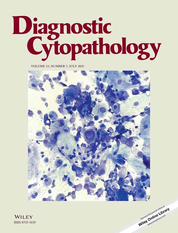
Tzanck smears, first introduced by Arnault Tzanck, are widely used in dermatology for the cytodiagnosis of cutaneous lesions. In the current era of evidence-based medicine with the availability of more confirmatory sophisticated tests, the use of this bedside tool is limited. In this article, the authors tried to assess the utility of Tzanck smear in various dermatological disorders. Findings in the Pemphigus group of disorders were studied in detail with complete and incomplete changes. Results were compared with histopathology, anti-desmoglein antibody status, and in combination. Further subtle features of the acantholytic cells were studied in detail. The sensitivity and specificity with histopathology and anti-desmoglein antibodies were 80.70%, 68.18% and 82.75%,100%. When patients were having either follow-up of histopathology or anti-desmoglein antibodies, each sensitivity was 81.01% and specificity was 87.50%. Complete acantholytic cells were predominant in 39.18%, and incomplete cells were predominant in 60.32% of cases. Rounding was the most consistent feature followed by nuclear enlargement. Streptocytes, dyskeratosis, and tadpole cells were also seen in a few cases. Other diagnoses of viral infections, like molluscum contagiosum (4 cases) and Herpes virus cytopathic effects (25 cases), were also observed in the study. Finally, Tzanck smear is still useful in dermatology, which can aid in rapid diagnosis and starting early treatment. Incomplete acantholytic cells, when seen in smear, help to pick up Pemphigus disorders even if all the classical features are not seen.
Conflicts of Interest
The authors declare no conflicts of interest.
Open Research
Data Availability Statement
Data is available from the authors on request.