Adenoid cystic carcinoma: A study of 19 cases of salivary and extra-salivary tumours diagnosed by fine needle aspiration cytology
Abstract
Background
Adenoid cystic carcinoma (ACC) arises at sites where seromucinous or sweat gland epithelium is present and commonly affects the salivary glands. Rarely it can occur at extra-salivary locations.
Methods
A retrospective analysis of 19 cases of ACC diagnosed on fine needle aspiration cytology (FNAC) over a period of 15 y (2002-2016) was made.
Results
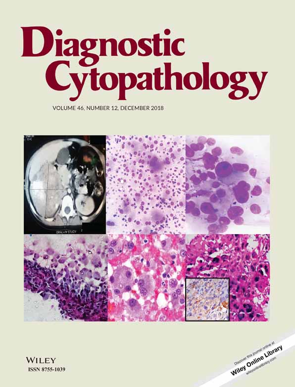
Of 19 total cases, there were 10 salivary and 9 extra-salivary ACCs. Extra-salivary tumours were seen in 2 cases each in trachea, tongue and in one case each in bronchus, lung, subcutaneous tissue, maxillary antrum, and external auditory canal. The age ranged from 14-80 y (mean: 49.5 y), 10 patients were male and 9 were female. The smears were highly cellular in 11 cases, moderately cellular in 5 cases while 3 cases were paucicellular. Multilayered dense clusters, tissue fragments, dispersed cells and cup-shaped fragments were seen. Relatively uniform cells with high nuclear: cytoplasmic ratio, hyperchromatic nuclei, irregular margins, and nuclear moulding were observed. Variable sized hyaline globules, finger-like hyaline material, hyaline cylinders, and hyaline cords were noted. The cytologic diagnosis of ACC was rendered in 13 cases while in 6 cases it was one of the differential diagnosis including monomorphic adenoma, membranous variant of basal cell adenoma, adnexal tumour, polymorphous adenocarcinoma, and pleomorphic adenoma (PA).
Conclusions
Cytologists must be aware of varied locations where ACC can occur. A diagnosis of ACC must not rely exclusively on the occurrence of hyaline globules but necessitates a close scrutiny of cellular and nuclear features to avoid diagnostic pitfalls.
CONFLICT OF INTEREST
Nil