Cytologic comparison of a primary parathyroid cancer and its metastatic lesions: A case report
Abstract
We describe the fine-needle aspiration cytology features of a primary parathyroid cancer and of the local recurrent and distant metastatic lesions. The presence of prognostic factors Ki-67 and proliferating cell nuclear antigen (PCNA) was compared immunohistochemically between primary parathyroid carcinoma and related metastatic and recurrent foci. Flow cytometric DNA analysis was also performed to investigate any chromosomal abnormality of the parathyroid carcinoma.
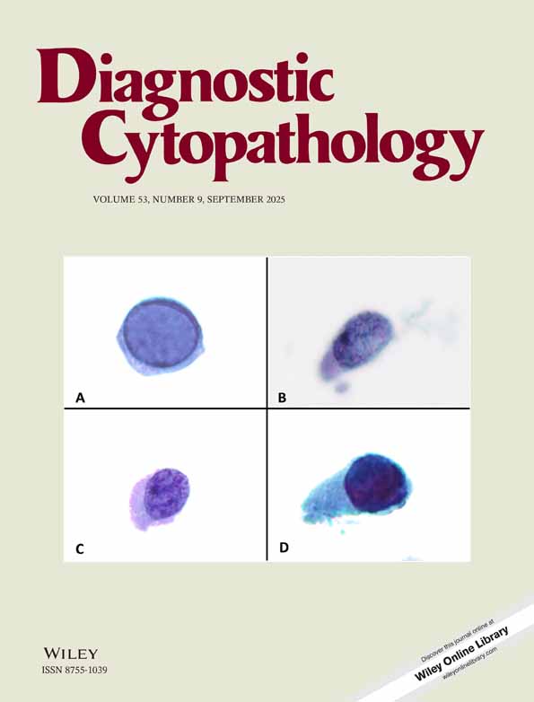
Cytologic examination of the endocrine tumor showed that it comprised a loose cohesive cluster and tumor cells with granular cytoplasm and mild nuclear atypia, but for purposes of cytodiagnosis, it is difficult to determine whether such a neoplasm is malignant on the basis of morphology alone. Immunohistochemical analysis showed that Ki-67 and PCNA labeling indices were higher in the recurrent and metastasized carcinomas than in the primary cancer, suggesting that neoplastic cells become more malignant in the recurrent and metastasized foci.
To our knowledge, this is the first report describing not only cytopathologic but also immunocytologic differences between primary parathyroid cancer and the metastatic lesion. Diagn. Cytopathol. 2006;34:50-55. © 2005 Wiley-Liss, Inc.