Volume 35, Issue 1 pp. 76-77
Erratum
Free Access

Erratum: Combined effects of 60 Hz electromagnetic field exposure with various stress factors on cellular transformation in NIH3T3 cells

Hae-June Lee

Hae-June Lee

Division of Radiation Effects, Korea Institute of Radiological and Medical Sciences, Seoul, Korea

Search for more papers by this author
Yeung Bae Jin

Yeung Bae Jin

Division of Radiation Effects, Korea Institute of Radiological and Medical Sciences, Seoul, Korea

Collge of Pharmacy & Division of Life Science & Pharmaceuticals, Ewha Womans University, Seoul, Korea

Search for more papers by this author
Jae Seon Lee

Jae Seon Lee

Division of Radiation Cancer Research, Korea Institute of Radiological and Medical Sciences, Seoul, Korea

Search for more papers by this author
Jong-Il Choi

Jong-Il Choi

Advanced Radiation Technology Institute, Korea Atomic Energy Research Institute, Jeongeup, Korea

Search for more papers by this author
Ju-Woon Lee

Ju-Woon Lee

Advanced Radiation Technology Institute, Korea Atomic Energy Research Institute, Jeongeup, Korea

Search for more papers by this author
Sung Ho Myung

Sung Ho Myung

Power Systems Research Division, Electrical Environment Team, Korea Electrotechnology Research Institute, Changwon, Korea

Search for more papers by this author
Yun-Sil Lee

Corresponding Author

Yun-Sil Lee

Collge of Pharmacy & Division of Life Science & Pharmaceuticals, Ewha Womans University, Seoul, Korea

Correspondence to: Prof. Yun-Sil Lee, College of Pharmacy & Division of Life Science & Pharmaceuticals, Ewha Womans University, 11-1 Daehyun-Dong Seodaemun-Gu, Seoul 120-750, Korea.

E-mail: [email protected]

Search for more papers by this author
First published: 08 October 2013

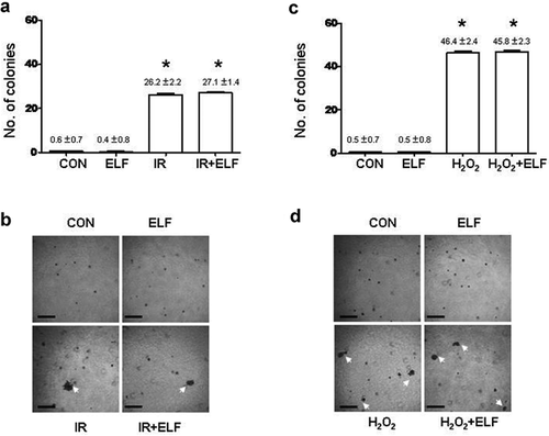
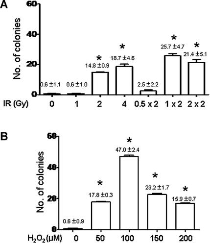
In the original article [Lee et al., 2012], the published Figure 3b is an image of Soft agar assay, but the same picture was uploaded to represent both IR and IR-ELF exposures. We have attached a corrected version of Figure 3. The scientific conclusions of the article were not affected by this error in anyway. The authors regret the mistake.
Details are in the caption following the image
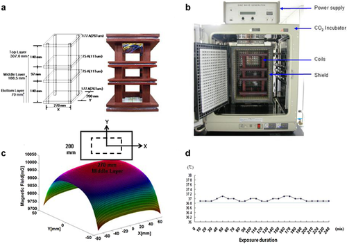
Schematic of the 60 Hz ELF magnetic field exposure system for in vitro studies. a: System structure of the magnetic field generator. b: Sixty hertz magnetic field exposure system included a power supply, magnetic field generating coils and a ferrite shielding system in a CO2 incubator. c: ELF magnetic field distribution for the middle floor of the exposure system. d: Maintenance of temperature in the exposure location during the exposure period.
Details are in the caption following the image
a: Anchorage-independent growth of NIH3T3 cells in soft agar after 4 h treatment of indicated concentrations of H2O2. b: Exposure to ionizing radiation (IR) with a single or fractionated dose. *Significantly different from untreated control cells at P < 0.05 (mean ± SD).
Details are in the caption following the image
a,b: Number of colonies with 4 h exposure of 1 mT ELF with or without two doses of 1 Gy IR. c,d: Treatment with 100 µM H2O2. b,d: Typical morphology of colonies after IR exposure or H2O2 treatment is also shown (white arrows). Scale bar = 50 µm. *Significantly different from untreated control cells at P < 0.05 (mean ± SD). CON, unexposed control cells; ELF, ELF-exposed cells; IR, ionizing radiation-exposed cells; H2O2, H2O2-exposed cells; ELF + IR, combined exposure of ELF and IR; ELF + H2O2, combined exposure of ELF and H2O2.
Details are in the caption following the image
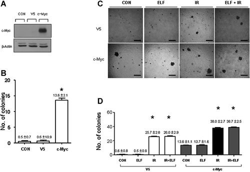
a: Imunoblotting of c-Myc protein after transfection of empty vector (V5) or c-Myc was performed in NIH3T3 cells. b: Cellular transformation assay in control and V5 or c-Myc transfected NIH3T3 cells. c: Morphologyand d: number of colonies with exposure to 4 h ELF (1 mT), 2 Gy IR, or ELF + IR in c-Myc transfected and V5 transfected cells. Scale bar = 50 µm. *Significantly different from corresponding untreated control cells at P < 0.05 (mean ± SD). CON, unexposed control cells; V5, pcDNA-V5 control plasmid transfected cells; c-Myc, c-Myc-V5 plasmid transfected cells; ELF, ELF-exposed cells; IR, ionizing radiation-exposed cells; ELF + IR, combined exposure of ELF and IR.

    The full text of this article hosted at iucr.org is unavailable due to technical difficulties.