Rapid airway stenosis due to ruptured occipital artery in a patient with neurofibromatosis type I
Abstract
Background
Neurofibromatosis type I is rarely associated with vascular abnormalities. Here, we report a case of rapid airway stenosis caused by a ruptured occipital artery that was treated with surgical airway management.
Case Presentation
A 40-year-old woman, with no medical history, presented with a chief complaint of a sudden neck pain on the left side. She had a prominent mass in the outer left side of the neck. After arrival at the emergency room, the patient complained of severe dyspnea and experienced a rapid drop in oxygen saturation. Supplemental ventilation was ineffective, and tracheal intubation was attempted; however, laryngeal expansion could not be observed because of the enlarged cervical mass. Therefore, to manage the surgical airway, a cricothyrotomy was first carried out, which resulted in an immediate increase in oxygen saturation. Two percutaneous embolizations and one surgical procedure were carried out, and the patient was discharged without any complications.
Conclusion
For a sudden onset cervical mass, airway management should be undertaken, keeping in mind the possibility of worsening rapid airway narrowing due to bleeding.
INTRODUCTION
Neurofibromatosis type I (NF-1), also known as von Recklinghausen disease, is an autosomal dominant inherited disorder caused by neuroectodermal tumors of the peripheral nervous system. The clinical manifestations of NF-1 are usually multiple café-au-lait spots, variable neurofibromas, Lisch nodules or iris hamartomas, freckles, abnormal bone lesions, pheochromocytomas, optic nerve gliomas, and Schwann cell tumors.1 Vascular abnormalities associated with NF-1 are rare and have been reported to occur in 0.4%–6.4% of patients.2 We report a case of rapid respiratory failure due to airway stenosis caused by a ruptured left occipital artery aneurysm that was successfully treated with surgical airway management.
CASE PRESENTATION
A 40-year-old woman presented with a chief complaint of a sudden pain on the left side of her neck while driving. She requested an ambulance and was transported to our hospital because of swelling in the left neck area as well as pain.
Her symptoms were as follows: Glasgow Coma Scale score, E4V5M6; body temperature, 37.1°C; blood pressure, 237/125 mmHg; pulse rate, 89 b.p.m.; respiratory rate, 25 breaths/min; and oxygen saturation in room air, 97%. There was a prominent mass on the outer left side of the neck, approximately 10 cm in diameter. There were café-au-lait spots all over the body and small scattered nodules. While preparing for a contrast computed tomography scan, the patient complained of severe dyspnea, expressed agony, and exhibited a rapid drop in oxygen saturation. She was immediately ventilated with a bag valve mask, but ventilation was impossible because her tongue had protruded from the oral cavity. When a laryngoscope was used to expand the laryngeal area with fentanyl 0.1 mg, 1% propofol 60 mg, and rocuronium 50 mg, the glottis could not be observed because it was compressed by the enlarged mass. Therefore, to manage the surgical airway, a cricothyrotomy was first carried out, which resulted in an immediate increase in oxygen saturation with a gaseous eruption. Subsequently, a gum-elastic bougie was inserted caudally and a 6 mm tracheal tube was inserted using the bougie as a guide.
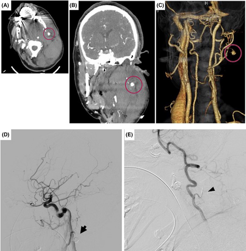
Contrast-enhanced computed tomography revealed a massive hematoma in the deep interstitial space from the left buccal region to the left side of the neck, which caused airway stenosis. There was a 1 cm-long aneurysm in the left occipital artery, with extravascular leakage around the aneurysm (Fig. 1A–C).

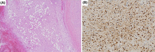
Angiography undertaken on the same day also showed a left occipital artery aneurysm, percutaneous embolization was selected based on café-au-lait spots and multiple nodules throughout the body, which suggested neurofibromatosis and vascular fragility. After embolization, we confirmed that the aneurysm was not contrasted and the procedure was completed (Fig. 1D). On day 2 of hospitalization, the swelling in the left side of the neck increased, and after an emergency angiogram was performed for suspected rebleeding, another coil embolization was performed. As the occipital artery was contrasted retrogradely from an artery branching from the vertebral artery to the vicinity of the aneurysm, we suspected bleeding around the aneurysm and undertook another embolization procedure. As the microcatheter could not be guided to the peripheral occipital artery, the branch from the vertebral artery was embolized with a coil at the bifurcation (Fig. 1E). However, due to the continued increase in cervical swelling, a left occipital artery aneurysmectomy was performed on day 5 of admission. From the time of skin incision, there was bleeding everywhere, and the total bleeding volume was 2,000 mL, making hemostasis difficult. When the sternocleidomastoid muscle was released from the mastoid process and rotated downward, a coil-filled aneurysm was exposed, which was dissected and removed together with the surrounding hematoma. The extraction sites were then examined pathologically, and the pathology results showed that it corresponded to a diffuse type of neurofibroma (Fig. 2). The patient was discharged without complications on day 31 since visiting the hospital.

DISCUSSION
According to the National Institutes of Health Consensus Development Conference Statement, NF-1 is diagnosed when two or more of six characteristic features are present.3 Although the frequency of NF-1 vasculopathy is reported to be rare, vasculopathy is the second most common cause of death in NF-1 patients, after malignancy.4 Vascular disorders of NF-1 include aneurysms, stenoses, and medium- and large-vessel arteriovenous malformations. The prevalent sites of aneurysms include the aorta, renal artery, mesenteric artery, carotid artery, and vertebral artery. Stenosis and aneurysms of the aorta, renal artery, mesenteric artery, and carotid artery are more likely to occur in younger patients than in older patients.2 To the best of our knowledge, there have been three previous case reports of airway emergencies due to rupture of the occipital artery caused by NF-1,5-7 but this is the rare case in which surgical airway management saved the patient's life (Table 1). In addition, none of the case reports of ruptured occipital artery aneurysms to date have presented pathology, and in this case, the pathology diagnosis of NF-1 was confirmed.
| Case | Sex | Age (years) | Author, year (ref. no.) | Airway management | Treatment procedure | Outcome |
|---|---|---|---|---|---|---|
| 1 | M | 52 | Takeda et al. 20025 | Intubation | Embolization | Died |
| 2 | F | 60 | Takeda et al. 20025 | No airway emergency | Embolization | Survived |
| 3 | M | 43 | Takeda et al. 20106 | Surgery | Embolization | Survived |
| 4 | M | 48 | Kanematsu et al. 20118 | Not mentioned | Embolization | Survived |
| 5 | F | 39 | Kanematsu et al. 20118 | No airway emergency | Embolization | Survived |
| 6 | F | 70 | Imahori et al. 20169 | No airway emergency | Embolization | Survived |
| 7 | F | 53 | Bissacco et al. 20187 | Intubation | Embolization | Survived |
| 8 | F | 83 | Illuminati et al. 201810 | No airway emergency | Surgery | Survived |
| 9 | F | 40 | Present case | Surgery | Embolization and surgery | Survived |
- F, female; M, male.
The occipital artery is a branch of the external carotid artery. It arises posteriorly at the same level as the facial artery, passes below the posterior belly of the digastric muscle, and extends toward the occipital region. Because of the anatomical proximity of the occipital artery to the trachea, a rupture can rapidly obstruct the airway. In the present case, it took 56 min from onset and 10 min from arrival at the hospital for a significant decrease in oxygen saturation to occur. Therefore, it is necessary to act in anticipation of sudden airway narrowing and to be prepared for surgical airway management, in addition to the usual airway securing methods. In particular, because tracheal deviation caused by an increasing hematoma could make even normal intubation difficult, it is important that intubation be carried out by the most skilled physician available and that someone capable of surgical airway management be prepared at the same time.
CONCLUSION
For a sudden onset cervical mass with a history of NF-1 or in situations where NF-1 is suspected, airway management should be performed taking into account the possibility of worsening rapid airway narrowing due to bleeding.
ACKNOWLEDGMENTS
We have reported our experience with a patient who received treatment at the emergency center of Ichinomiya Nishi Hospital. We thank all physicians and staff members of the hospital.
DISCLOSURE
Approval of the research protocol: N/A.
Informed consent: The patient provided consent for publication.
Conflict of interest: None.


