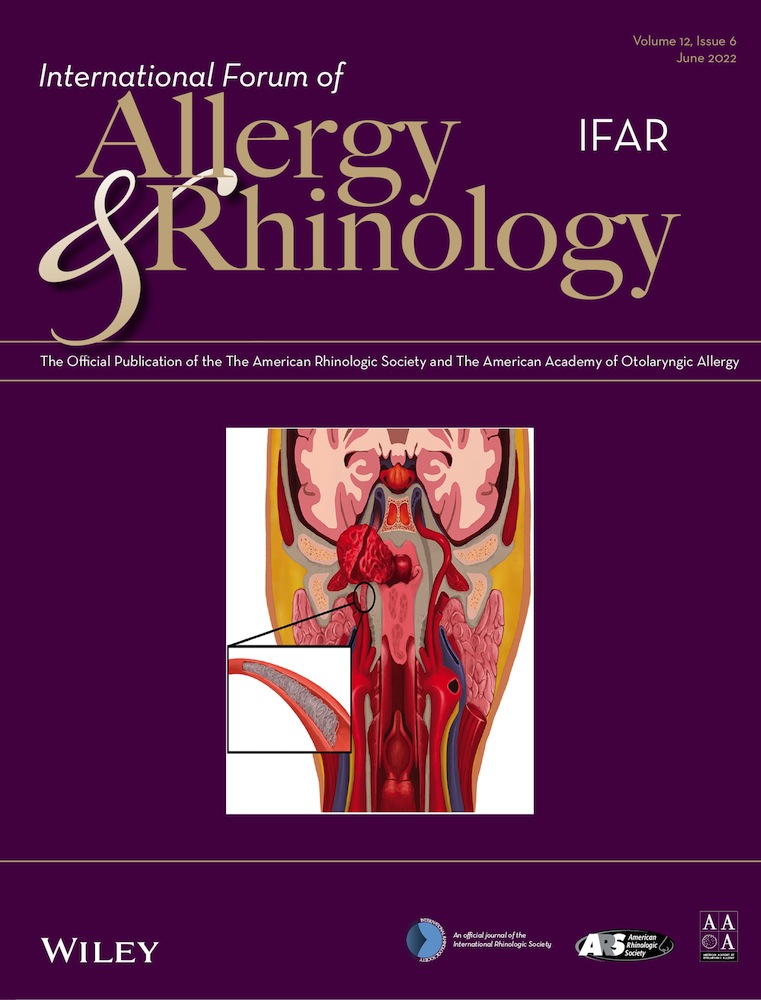
Usefulness of imaging studies for diagnosing and localizing cerebrospinal fluid rhinorrhea: A systematic review and meta-analysis
Additional Supporting Information may be found in the online version of this article.
Potential conflict of interest: None provided.
The English in this document has been checked by at least two professional editors, both native speakers of English. For a certificate, please see: http://www.textcheck.com/certificate/hK0p97
View this article online at wileyonlinelibrary.com.
Abstract
Background
Our aim in this study was to determine the usefulness of diagnosis by imaging studies for the localization of cerebrospinal fluid (CSF) rhinorrhea.
Methods
PubMed, SCOPUS, Embase, Web of Science, and Cochrane Library databases were searched up to July 2021. True and false positive and negative data were collected along with the characteristics of each study. Methodologic quality was assessed using the QADAS-2 tool.
Results
Sixteen studies involving 472 patients were included. The diagnostic odds ratio (DOR) of imaging studies was 13.6195 (95% confidence interval [CI], 7.4756-24.8129; I2 = 28.1%). The area under the summary receiver-operating characteristic curve was 0.712. Sensitivity, specificity, negative predictive value, and positive predictive value were 0.8507 (0.7773-0.9029), 72.1%; 0.7827 (0.6865-0.8556), 26.8%; 0.5828 (0.4398-0.7132), 67.4%; and 0.9407 (0.8935-0.9678), 59.1%, respectively. In the subgroup analysis, there were significant differences for sensitivity (computed tomography [CT], 0.7421; computed tomography cisternography [CTC], 0.8872; magnetic resonance imaging [MRI], 0.8365; magnetic resonance cisternography [MRC], 0.8565; intrathecal gadolinium magnetic resonance cisternography [GaMRC], and 0.9307; radionuclide cisteronography [RNC], 0.7097; p = 0.0481) and for negative predictive value among imaging modalities (CT, 0.3028; CTC, 0.4848; MRI, 0.4658; MRC, 0.7465; GaMRC, 0.8611; and RNC, 0.5263; p = 0.0046). There were no significant differences among imaging modalities for specificity, positive predictive value, or DOR (p > 0.05).
Conclusion
Imaging studies can be used in the diagnosis of CSF rhinorrhea. Gadolinium magnetic resonance cisternography showed the highest diagnostic accuracy. MRC showed fair diagnostic accuracy without intrathecal injection.


