Predicting the Position of Hip Bones Within the Pelvic Girdle: A Case Study of the Kebara 2 Neanderthal
Funding: This work was supported by the University of California, Davis, Department of Anthropology.
ABSTRACT
Objectives
Poor preservation of hominin pelvises and the lack of soft tissue in the fossil record inhibits researchers' abilities to ascertain the true geometry of hominin pelvic girdles. The reconstruction process becomes subjective, largely relying on researchers' anatomical expertise, particularly, when the sacrum is absent or cannot be used to orient the hip bones. The bilateral symmetry of the pelvis, however, offers an opportunity to use one side to reconstruct potentially missing data on the other side.
Methods
We developed a regression model to predict the translation and rotation actions that are needed to transform a hip bone onto the location of its pair. We collected landmarks and curve semilandmarks on a training sample of medical CT scans of 103 adult humans. A reduced rank regression model was trained to predict the values that would fit each right hip bone on its left pair. Then, we applied the model to two reconstructions of the Kebara 2 Neanderthal pelvis and assessed how well, it predicted the reconstructions (assuming the sacrum was absent), which were made using the preserved sacrum.
Results
Euclidean errors from the model were significantly lower than errors from a mean form model and an observed form pairwise model.
Conclusion
Regression modeling that takes advantage of bilateral symmetry can be used to reliably predict “missing” human hip bones and Kebara 2's reconstructed left hip bones. This method can be employed in conjunction with a researcher's anatomical expertise and other techniques to reduce subjectivity in the fossil pelvis reconstruction process.
1 Introduction
The shape of the bony pelvis has undergone multiple changes through human evolutionary history, and these changes have been shaped by and have impacted childbirth (Arsuaga et al. 1999; Gruss and Schmitt 2015; Huseynov et al. 2016; Kappelman 1996; Rosenberg 1992; Rosenberg and DeSilva 2017; Rosenberg and Trevathan 2002; Ruff 1995, 2010; Simpson et al. 2008; Stansfield et al. 2021; Tague and Lovejoy 1986; Trinkaus 1984; Walrath 2003; Weaver and Hublin 2009), thermoregulation (Holliday 1997; Rosenberg et al. 2006; Ruff 1991, 1994, 1995, 2010; Weaver and Hublin 2009), posture, locomotion (Bailey et al. 2019; Been et al. 2013, 2017; Lovejoy et al. 2009; Lovejoy 2005; Middleton et al. 2017; Rak 1993; Stern et al. 1983), and other biological processes related to the pelvis. Further investigations of these factors and their impacts on the geometry of the pelvic girdle are dependent on accurate reconstructions.
1.1 Pelvic Reconstruction Challenges
In studies of the fossil record for human evolution the poor preservation of pelvic bones coupled with the absence of soft tissue inhibits researchers' abilities to ascertain the true spatial relationships between the bony elements of the pelvis in extinct hominins. Researchers have generally relied on the presence of a well-preserved sacrum that can be articulated confidently with what remains of the hip bones. Many of these reconstructions have had to utilize the bilateral symmetry of the pelvis to mirror a reconstructed hemipelvis about the midsagittal plane to form a full pelvic girdle (e.g., Adegboyega et al. 2021; Berge and Goularas 2010; Claxton et al. 2016; Rak and Arensburg 1987). In the absence of a sacrum, Weaver and Hublin (2009) fit together the right and mirrored left-sided fragments by matching overlapping anatomy to create a more complete right hip bone and then estimated sacral and other missing anatomical landmarks before also mirroring the hemipelvis to create the left side. In these cases, either a sacrum or an estimated sacrum was used to position the hip bones.
When the pubic symphysis is not preserved, a great amount of care must be taken in the orientation of the hip bones at the sacroiliac joint, as there is no other way of independently confirming that the hip bones have been angled correctly without the anterior constraints provided by the pubic symphysis (Claxton et al. 2016; Li 2002; Torres-Tamayo et al. 2023). These issues are well highlighted in Adegboyega et al. (2021) where even in the face of a relatively well-preserved sacroiliac joint, the pubic symphysis uncertainty contributed to differences in the reconstructions and morphological interpretations of the Kebara 2 Neanderthal pelvis. In many cases, however, the sacroiliac joint cannot be relied upon at all, either due to the poor preservation of the bones or to the complete absence of the sacrum. Under these conditions, the placement of the hip bones falls entirely on the estimated shape and size of the pubic symphysis which researchers use to complete the inlet anteriorly. Minor changes in orientation at the pubic symphysis however can drastically change the orientation of the hip bones, and this could have implications for our understanding of fossil hominin biology (Häusler and Schmid 1995; Lovejoy 1979; Schmid 1983; Tague and Lovejoy, 1986). Furthermore, reported pubic symphysis dimensions vary widely from study to study (see Becker et al. 2010, 2014). Now, using the method we present below, we provide an independent check on pelvic reconstructions carried out when the sacrum is present, but the pubic symphysis is poorly preserved.
1.2 The Kebara 2 Neanderthal Pelvis
The Kebara 2 Neanderthal is a Levantine Neanderthal partial skeleton dated to 60–65 ka (Valladas and Valladas 1991). The skeleton, which is presumed to have belonged to an adult male, has been crucial to our understanding of Neanderthal postcranial anatomy. For example, it has been used to represent Neanderthal body breadth and pelvic canal shape which in turn have been used to understand Neanderthal climatic adaptation, birth mechanism, infant head size, and other biological processes (Adegboyega et al. 2021; Been et al. 2010, 2017; Chapman et al. 2017; García-Martínez et al. 2014; Gómez-Olivencia et al. 2009, 2013, 2017, 2018; Rak and Arensburg 1987; Torres-Tamayo et al. 2020; Vandermeersch 1991). The pelvis consists of a left hip bone which is missing the pubis as far back as the root of the iliopubic ramus and the entire ischiopubic ramus, a right hip bone which is relatively well preserved, and a sacrum which is essentially complete although exhibiting some developmental anomalies and postmortem damage (Duday and Arensburg 1991; Rak and Arensburg 1987; Trinkaus 2018).
The Kebara 2 pelvis has undergone several reconstructions using both physical and virtual methods. The first reconstruction by Rak and Arensburg (1987; see also Rak 1991) was achieved by articulating a cast of the right half of the sacrum with a cast of the right hip bone to form a hemipelvis. The missing region of the pubis was reconstructed following the curvature of the Linea terminalis (i.e., the inlet rim) so the full pelvis was only visible when physically mirror imaged. Sawyer and Maley (2005) used the sacrum, right ilium, and ischium of Kebara 2 with the left ilium and ischium from Feldhofer 1, and the pubis from La Ferrassie 1 to build a complete pelvis, but the composite nature of the reconstruction and the artistic license employed “in order to maintain symmetric continuity” make it difficult to assess (Torres-Tamayo et al. 2020). Adegboyega et al. (2021) applied virtual and physical reconstruction techniques to produce two reconstructions of the Kebara 2 pelvis. Virtual techniques were applied independently by two researchers to surface renderings generated from computed tomography (CT) scans to realign misaligned fragments of the hip bone and sacrum based on the same general reconstruction protocol and each researcher's assumptions about how the fragments should be aligned. These techniques also allowed the researchers to create mirror images of the right hip bone and the right half of the sacrum to create a new left side to replace the poorly preserved left side. Physical techniques were then used to articulate three-dimensional (3D) printouts of the reconstructed elements which in turn were used to articulate the virtual elements using landmark data and image warping techniques to create two articulated virtual pelvic girdles that matched the physical replicas. Presenting more than one reconstruction allowed for the assessment of the assumptions and choices made by different researchers and how they impact the reconstructions. This process yielded two reconstructions with the most noticeable differences in the shape and size of the pelvic outlet. Some of these differences can be attributed to independent choices made by each researcher when aligning the bone fragments; however, some of them are also the result of different choices that were made when orienting the hip bones at the sacroiliac joint. This example illustrates the need for more systematic methods that could be used to support researchers' anatomical expertise and reduce uncertainty in fossil pelvis reconstructions.
1.3 Missing Data Estimation and Bilateral Symmetry
Both physical and virtual techniques have been employed in previous pelvis reconstructions. Virtual reconstruction methods have become more popular due to their ability to minimize the risk of damage to already fragile fossil elements (Gunz et al. 2009; Weber and Bookstein 2011; Zollikofer et al. 2005) and to readily create multiple reconstructions (Gunz et al. 2009; Adegboyega et al. 2021). 3D geometric morphometrics (3DGM) has played an important role in virtual reconstruction as a way to estimate missing data (Benazzi et al. 2009; Brassey et al. 2018; Gunz et al. 2009; Gunz and Mitteroecker 2013; Mitteroecker and Gunz 2009; Schlager et al. 2018; D. Slice 2005; D. E. Slice 2007).
There are two main 3DGM methods for missing data estimation, which in principle could be used to estimate the form and orientation of the hip bones. One is using a thin-plate spline to predict the coordinates of missing landmarks by deforming a reference individual to the target individual with missing landmarks (Bookstein 1989, 1991; Gunz et al. 2005; Mitteroecker and Gunz 2009). The other is regression, which works by regressing variables (e.g., 3D coordinates) collected on the element to be reconstructed on other variables in a training sample with the complete set of landmarks, so that the missing coordinates from the element to be reconstructed are predicted by the regression model (Bookstein et al. 2003; Gunz et al. 2004; Stelzer et al. 2018; Torres-Tamayo et al. 2020; Weber and Bookstein 2011). Neither of these methods, however, explicitly make use of bilateral symmetry. Our goal was to develop a method that explicitly took advantage of the bilateral symmetry of the pelvis.
1.4 Study Aims
We investigate whether the bilateral symmetry of the pelvis can be used to accurately predict the form and position of a hip bone from the hip bone of the other side. Specifically, we use reduced rank regression (RRR) to predict the translations and rotations needed to transform a hip bone onto the location of its pair. The novelty of our approach is that unlike existing missing data estimation methods used in fossil hominin reconstructions in which the set of 3D coordinates of the missing elements are predicted explicitly, we predict instead the translations and rotations that transform one hip bone to its symmetric counterpart. We thus obtain the coordinates of the missing side implicitly. After evaluating the approach in living humans, we applied it to the two reconstructions of the Kebara 2 Neanderthal pelvis presented in Adegboyega et al. (2021).
2 Materials and Methods
2.1 Sample and Medical Images
Data were collected from 103 adult humans (n = 52, males; n = 51, females) ranging between the ages of 20 and 96 years old, who had undergone abdominal, and pelvis CT imaging between May 2019 and November 2020 through the University of California Health System in California, USA (Table 1). The training sample was intentionally selected to include a wide range of ages and demographic characteristics to evaluate the method's robustness to variation. We obtained approval from the University of California, Davis, Institutional Review Board (Davis, CA, USA; protocol 1100046-1) to use these patients' data in this study. The scans were taken using one of the following medical CT scanners at slice thicknesses ranging between 1 and 1.5 mm: Siemens Somatom Definition DS 64, Siemens Somatom Definition AS 128, Siemens Somatom Sensation PCH 64, and GE Light Speed VCT 64. Individuals who presented with injuries including but not limited to pelvic fractures, neuromuscular disorders, and pubic symphysis diastasis, or who were identified as having undergone treatments in the past for pelvic injuries were not included in the study as these conditions might influence their pelvic morphology.
| Min | Max | Mean | SD | |
|---|---|---|---|---|
| Age (years) | 20.0 | 96.0 | 54.3 | 20.6 |
| Weight (kg) | 44.7 | 146.8 | 77.6 | 21.1 |
| Height (m) | 1.47 | 1.93 | 1.67 | 0.10 |
- Abbreviation: SD, standard deviation.
The CT scans were imported as DICOM (.dcm) files into Avizo Lite 9 (Thermo Fisher Scientific, Waltham) for segmentation and data collection. The skeletons were first segmented from soft tissue using automated density thresholding tools; then, the pelvic bones (the left and right hip bones and the sacrum) were manually segmented from the skeletons. Finally, 3D surface renderings were generated in Avizo for each segmented pelvis to be used in the analyses. To test the predictive model on a pelvis from a hominin taxon other than Homo sapiens, we used the surface renderings of the two Kebara 2 Neanderthal pelvis reconstructions reported in Adegboyega et al. (2021).
2.2 Landmarks and Semilandmarks
3D landmarks and semilandmarks were collected on each pelvis in Avizo. A set of anatomical landmarks (Dryden and Mardia 1998) were manually collected on the left and right hip bones. Curve semilandmarks were collected along the inlet rims of the left and right hip bones, as well as along the margins of the articular surfaces of the bony pubic symphyses. These landmarks and semilandmarks represent the forms and, because the pelvis is articulated, positions of the hip bones within the pelvic girdle. To ensure roughly equidistant homologous points for each individual (Gunz et al. 2005), the semilandmark sets were resampled using the function equidistantCurve from the R package Morpho v 2.11 (R Core Team 2023; Schlager 2017). The final count of semilandmarks was 72; 22 along the inlet rim of each (right and left) hip bone, and 14 along each pubic symphysis (Figure 1; Table 2).

| Landmark | Definition |
|---|---|
| 1 | Apex of the anterior superior iliac spinea |
| 2 | Most superior point on the iliac crest |
| 3 | Apex of the posterior superior iliac spinea |
| 4 | Apex of the posterior inferior iliac spinea |
| 5 | Most inferior point of the arcuate line of the ilium |
| 6 | Midpoint of the superolateral edge of the cristal tubercle |
| 7 | Deepest point of the greater sciatic notch |
| 8 | Point where the iliopubic ramus meets the arcuate line |
| 9 | Apex of the ischial spine |
| 10 | Most superior point of the ischial tuberosity |
| 11 | Most inferior point of the ischial tuberosity |
| 12 | Most superior point of the medial aspect of the pubic symphysis |
| 13 | Most inferior point of the medial aspect of the pubic symphysis |
| 14 | Most anterior point of the pubic tubercle |
| 15 | Most anterior point of the obdurator foramenb |
| 16 | Most superior point of the obturator foramenb |
| 17 | Most posterior point of the obturator foramenb |
| 18 | Point on the acetabulum margin corresponding to where ilium and iliopubic ramus meetb |
| 19 | Point on the acetabulum margin furthest away from landmark 18 |
| 20 | Most inferior point on the anterior lunate surface of the acetabulum margin |
| 21 | Point on the acetabulum margin furthest away from landmark 20 |
| 22 | Deepest point of the acetabular fossa |
| 23–36 | Curve semilandmarks along the pubic symphysis |
| 37–58 | Curve semilandmarks along the Linea terminalis of the hip bone |
2.3 Predicting Hip Bone Position
Our goal was to develop a systematic method for orienting fossil hominin hip bones within the pelvic girdle, to complement reconstructions created through more subjective methods. We used the bilateral symmetry of the pelvis to constrain the scope of our predictions, on the assumption that the right and left hip bones are approximately mirror images of one another. The training set of complete pelvises provided information about how right and left sides relate to each other through the actions of rigid translations and rotations. Using a statistical procedure, we inferred these actions from the training set and applied the model to predict the position of the reconstructed left hip bone of the Kebara 2 pelvis, based on its better-preserved right side. While we chose to predict the left side from the right, the code could be applied to predict the right side from the left just as easily.
We conducted our analysis in two stages, first training and testing the model on the human sample, and then applying the fitted model to the Kebara 2 pelvic reconstructions from Adegboyega et al. (2021). We began by registering the pelvises of the training set based only on the right hip bones (the predictors) via generalized procrustes analysis (GPA; procSym, Morpho v 2.11; Schlager 2017). This ensured that the data of the left hip bones (the targets) did not influence the Procrustes fitting, to mimic a scenario whereby only one hip bone is available. GPA translates all configurations of (semi-) landmarks to the origin, scales them to unit centroid size, and rotates them around the origin to minimize the total sum of squared distances between them (Dryden and Mardia 1998; Gower 1975; Zelditch et al. 2012). During the GPA, the semilandmarks were slid to minimize the bending energy between each individual and the Procrustes mean shape (Gunz et al. 2005). The centroid sizes for each right hip bone were stored for later rescaling.
2.3.1 Preparing the Predictors and Targets
To prepare the predictors and targets, we multiplied each Procrustes-registered pelvis by the centroid size of its right hip bone, which was the scale factor for the GPA. We then obtained the transformations that would best fit the right hip bone onto the left hip bone within each pelvic girdle using the function rotonto, which minimizes the sum of squared errors of the fit (Morpho v 2.11; Schlager 2017). The transformations of interest were the following: (1) reflection: creating a mirror image of the right hip bone; (2) translation: moving the mirrored hip bone to the position of the observed left hip bone; and (3) rotation: turning the reflected and translated hip bone to fit the orientation of the left hip bone (Figure 2).

where the reflection is about the x axis. The translation returned by rotonto is a 3 × 1 vector giving the linear shift between the predictor and the target. The rotation is a 3 × 3 matrix used to rotate a rigid body in Euclidean space. We reduced the rotation matrices to 3 Euler angles (e.g., Weaver et al. 2014) using rot2eul (Directional v 6.2, Tsagris et al. 2023) which, when combined with the translations, produced a set of six unique transformations for each individual. Our premise is that the 3D coordinates of the right hip bone contain statistical information about the six derived transformations. Thus, our initial modeling objective is to infer the transformations from the right hip bone coordinates.
2.3.2 Predicting the Left Hip Bones in the Training Set
A statistical model treating the right hip bone coordinates as covariates and the six transformations as outcomes produces a “many-to-many” prediction that, additionally, implies a reduction in dimension, as the number of right hip bone coordinates is much larger than the number of transformations. We chose a RRR model (Izenman 1975), related in concept to the estimation of canonical variates, for this purpose.
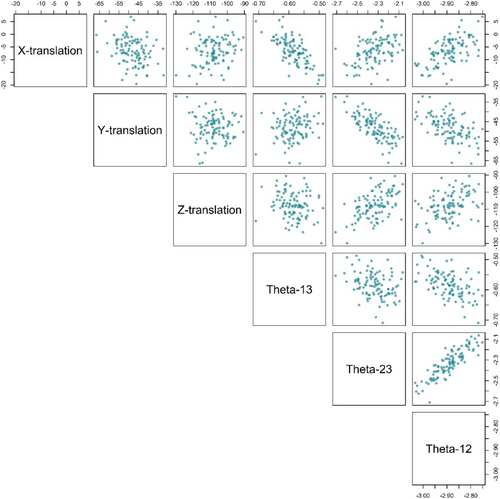
To determine the rank of our RRR model, we examined a pairwise scatterplot matrix displaying the covariation of the six transformations in the training sample. We additionally calculated the singular values of the matrix of sample transformations. Scatterplots exhibiting ample pairwise variation, and singular values bounded above zero, would indicate that the matrix of transformations is of full rank and represents six distinct dimensions.
A preliminary step in fitting an RRR model was to center and scale the reflected right hip bone coordinates and transformations so that all the values had a mean of zero and a standard deviation of 1. This prevented numerically larger coordinates and transformations from having oversized influence on the reduced-rank solution.
We predicted the six transformations and thus the left hip bone coordinates in the training set using a leave-one-out (LOO) procedure, in which each individual is held out of the sample, in turn. Under the LOO procedure, an RRR model was fitted to the remaining individuals, and the model coefficients were used to predict the outcome of the held-out individual. Here, the initial outcome consisted of the six transformations; from these, we obtained the individual's predicted left hip bone coordinates. The LOO procedure mimics a situation in which the left hip bone is incomplete or missing and the right hip bone coordinates are used to predict the left. We obtained a mean squared prediction error (MSE) for each individual in the training sample, defined as the Euclidean distance between the observed and predicted left hip bone coordinates, as part of the LOO procedure.
2.3.3 Predicting the Kebara 2 Reconstructions' Left Hip Bones
The RRR model can be used to assess the two reconstructions of the Kebara 2 pelvis presented in Adegboyega et al. (2021), through a comparison of the reconstructed left hip bones with their RRR predictions. Because Kebara is missing some of its pubic bone, we used the thin plate spline interpolation (Bookstein 1989, 1991) to impute its missing landmarks and semilandmarks using the human sample as a reference. Given that much of the pubic bone is preserved, we expect the thin-plate spline method to provide an accurate reconstruction of the missing portions (Gunz et al. 2009).
We then slid Kebara's semilandmarks and then, its newly constituted coordinate data were Procrustes registered onto the human sample using only the right hip bone landmarks, as was done with the human training sample. We then predicted the Kebara 2 left hip bone for each reconstruction using a RRR model fitted to the modern human sample, as in the LOO procedure.
2.3.4 Assessing Model Performance
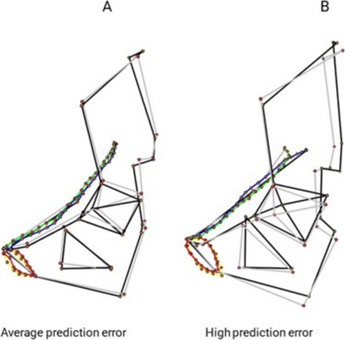
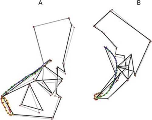
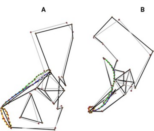
To assess how well the model predicted left hip bone coordinates, we generated wireframe contrasts depicting the observed and predicted hip bones from the human training sample, as well as predicted and reconstructed Kebara 2 hip bones. We report a selection of the human contrasts, as well as contrasts for both Kebara 2 reconstructions.
- LOO prediction errors from the RRR model for each individual's left hip bones in the training sample.
- Prediction errors from the mean form of the left hip bones in the training sample.
- Pairwise Euclidean distances between the coordinates of all the left hip bones in the training sample.
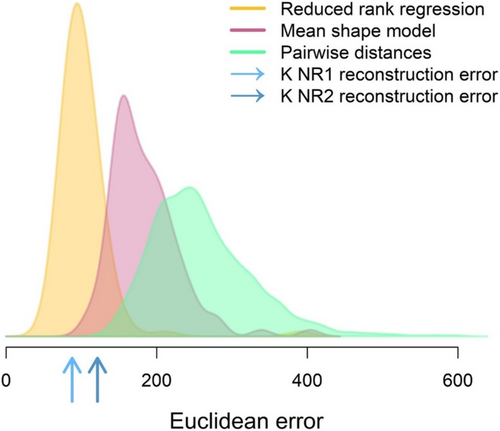
We summarized each of the three measures of form difference across the sample in a density plot and superimposed them for comparison.
The Kebara 2 reconstruction errors are sums of squared deviations between the RRR predictions and the reconstructed left hip bones, producing a single value for each of the two reconstructions. We marked these values on the error density plots for comparison.
All geometric morphometric analyses were carried out using the following packages in R: Morpho v 2.11 (Schlager 2017), geomorph v 4.0 (Adams et al. 2021; Baken et al. 2021), Directional v 6.2 (Tsagris et al. 2023), rrpack v 0.1–11 (Chen and Wang 2017), and shapes v 1.2.6 (Dryden 2021).
3 Results
3.1 Comparison of Predictions With Observed Hip Bones
The pairwise scatterplot matrix of transformations (Figure 3) shows variation in each of the six transformation parameters, with moderate correlation between some parameter pairs. The smallest singular value of the sample matrix of transformations, d6 = 0.29, is securely above zero, indicating that six identifiable dimensions are present. Figure 4 depicts the wireframe contrasts of the observed and predicted left hip bones of two individuals in the human training sample (see also the Supporting Information for interactive plots and triangular mesh files illustrating the contrasts). Figure 4A shows the results for a randomly selected individual, chosen from those with prediction errors close to the mean. For this individual, the model very reliably predicted the location of the hip bone; however, the predicted hip bone is slightly more medially positioned compared to the observed hip bone. This difference is most clearly highlighted at the acetabulum and the inlet rim. Figure 4B on the other hand depicts the difference between the observed and predicted hip bone for a randomly selected individual with a high prediction error (i.e., an individual in the fourth quartile of prediction errors). For this individual, the predicted hip bone still overlays the observed left side, but it is more anterolaterally positioned than the observed hip bone.
Figures 5 and 6 (see also the Supporting Information) show the contrasts of Kebara 2 reconstructions from a lateral view (Figure 5a & 6a) and a superior view (Figure 5b & 6b). In Figure 5 the prediction for reconstruction 1 (R1) lies almost entirely on the observed coordinates; however, the prediction is slightly tilted upwards in the sagittal plane from the center so that the posterior ends of the obturator foramen and the acetabulum are more anteriorly positioned. The difference between reconstruction 2's (R2) prediction and its observed configuration is more pronounced (Figure 6). Though the two configurations still overlap each other almost entirely, there is a greater distance between them which is highlighted by the positions of their ilia and the rotation of their acetabulae. The predicted hip bone is more anterosuperiorly positioned and more medially rotated than the observed hip bone.
3.2 Comparison of Prediction Errors
We created and plotted error densities to compare form deviations of different types (Figure 7). The plot shows that the RRR model performs favorably (LOO MSE = 102.52) in comparison to the mean form model (MSE = 183.71) and the set of all pairwise distances (mean squared deviation = 257.79). The reconstruction errors (R1 = 87.74; R2 = 121.27) for Kebara 2 fall within the bulk of prediction errors for the training set, slightly above the modal error.
4 Discussion
In this study, we applied a RRR model to predict the translation and rotation values needed to transform a human right hip bone to the left side. By also testing the method on the Kebara 2 pelvis, we showed that this method can be used to predict the hip bone position of a Neanderthal pelvis and potentially the hip bones of other members of the genus Homo. In this section, we further discuss the results of this study along with its limitations; the validity of the human training sample; the novelty of the method; and implications for assessing the Kebara 2 and future pelvic reconstructions.
4.1 Novelty of the Method
The two main 3DGM methods for missing data estimation—thin-plate spline and multivariate regression—both aim to predict the coordinates of each of the landmarks characterizing the missing part (Gunz et al. 2004, 2009; Stelzer et al. 2018; Torres-Tamayo et al. 2020). Here, instead of predicting numerous landmark coordinates, we predict only six values that describe the actions needed to create a bilaterally symmetric left hip bone from a reflected right hip bone. By making use of the bilateral symmetry of the pelvis, our approach substantially reduces how many quantities need to be predicted and uses the relative positions of the landmarks on the preserved side to constrain the relative positions of the landmarks on the predicted side. This is similar conceptually to reflecting a partial cranium and fitting the reflection to the original (Gunz et al. 2009), except that our approach does not rely on having overlapping anatomy in the original and reflection. The six values included the 3 × 1 translation matrix to move an already reflected right hip bone over to the left side, and the 3 Euler angles that oriented the new left side on the observed form, minimizing the sum of squared errors between them. The six actions, along with the coordinates of the reflected right hip bone, are sufficient to reconstitute a new left side.
4.2 Validity of the Predictors
The human training sample used in this study was randomly selected from patient CT scans that were acquired from University of California Health System medical records. We did not control for population diversity; however, according to the U.S. Census Bureau (2021), the 2020 US Census, self-reported racial and ethnic makeup of California was, Hispanic or Latino alone 39.4%; Non-Hispanic White alone 34.7%; Black or African American alone 5.4%; American Indian and Alaska Native alone 0.4%; Asian alone 15.1%; Native Hawaiian and Other Pacific Islander alone 0.4%; Some Other Race alone 0.5%; Two or More Races 4.1%. Because this was the pool from which our sample was collected, we are confident that the individuals reflect a broad range of population histories and thus we have accounted for a significant portion of pelvic form variation.
Nonetheless, this study is based on predictive models that were trained exclusively on living humans, therefore, an implicit assumption of our approach is that Neanderthals and living humans had similar relationships between hip bone form and positioning. While Neanderthal and human pelvises share many traits, the Neanderthal pelvis retains more ancestral traits, such as more flared iliac blades, a mediolaterally wider inlet, and longer pubic bones (Adegboyega et al. 2021; Arsuaga et al. 1999; Ponce de León et al. 2008; Rak and Arensburg 1987; Rosenberg 2007; Weaver and Hublin 2009). These morphological differences could be associated with different relationships between hip bone form and positioning. Nevertheless, our model predictions for Kebara 2 were largely consistent with the reconstructions made by Adegboyega et al. (2021) who used the sacrum to position the hip bones, suggesting that if there were different relationships in humans and Neandertals, the differences were not great enough to bias predictions based on Neandertal hip bones. To expand our approach for making predictions for a wider range of hominin taxa, for example, early hominins like Ardipithecus or Australopithecus, the inclusion of a variety of extant hominids might be necessary, as a human training sample alone might not be sufficient for characterizing the relationships between hip bone form and positioning (Lovejoy et al. 2009; White et al. 2015). Additionally, we would need to evaluate the model's performance when large sections of the hip bone are missing and must be reconstructed as an initial step.
We selected the Kebara 2 pelvis for this study because unlike other Neandertal pelvic remains that require extensive reconstruction, e.g., Tabun C1 (Ponce de León et al. 2008; Weaver and Hublin 2009), the well preserved Kebara 2 retains a substantial portion of the anatomy. This means that the uncertainty that is commonly associated with pelvis reconstruction is reduced with Kebara 2, making it a suitable fossil individual to test this predictive method.
4.3 Assessing Model Performance
The results of our study show that within humans and Neanderthals it is possible to accurately predict the positioning of hip bones within a pelvic girdle by transforming a “present” side as a replacement for the “missing” side. When we compared the Euclidean errors from our model to errors from a mean form model and an observed form pairwise model, the errors from the RRR model were notably lower. We were able to observe this visually with the wireframe contrasts which showed that in both the human sample and Kebara 2 the model positioned and oriented the predictions with very little error. Because the pelvis is not perfectly bilaterally symmetrical, some of the prediction error being captured consists of differences between the sizes and shapes of the left and right sides. We considered symmetrizing the pelvises in the training sample, which would have reduced the prediction errors; however, we thought it was important to report the more realistic expectation of this method to highlight the natural asymmetry that is lost in the mirroring process of most pelvic reconstructions, and thus capture the total error expected when this method is applied in a real fossil context. We also noticed that the pairwise comparison between the predicted and observed coordinates for Kebara 2 yielded a lower prediction error for R1 (87.74) than for R2 (121.27). Furthermore, when compared to the human mean prediction error (102.52), the prediction error for R1 was closer to the mean than for R2. This disparity might be the result of the different anatomical assumptions that were used to create each reconstruction. It is possible that the lower prediction error for NR1 means that NR1 should be preferred, but because we do not have the true Kebara to compare to, we hesitate to draw this conclusion. What we can say is that the R1 reconstruction conforms more closely than the R2 reconstruction to the model's predicted form.
As was mentioned above, this method has the potential to be applied more broadly to other hominin pelvises and to other bilaterally symmetric anatomical structures given that good reference samples are selected, and the associations and covariations between those structures are well understood (Gunz et al. 2009; Stelzer et al. 2018; Torres-Tamayo et al. 2018). If applied appropriately, predictive methods such as the one described here can provide statistical support to more manual or subjective reconstruction processes thereby reducing interobserver error in fossil pelvis reconstructions.
5 Conclusion
This work contributes to the growing application of 3DGM for predicting missing aspects of fossil morphology. In this study, we were able to use RRR analysis to recreate “missing” hip bones within our human sample, but even more encouragingly, the model was also able to make accurate predictions for a Neanderthal. The method we have proposed here aims to reduce subjectivity in hominin pelvis reconstructions through the application of statistical predictions, to systematically constrain the placement of hip bones even without the presence of a sacrum or other anatomical references, and to reduce the reliance of researchers' individual anatomic presumptions. We would like to emphasize that this method does not replace physical or other virtual reconstruction techniques, but rather, it can help to support these other methods by constraining areas of uncertainty.
Author Contributions
Mayowa T. Adegboyega: conceptualization (equal), data curation (lead), formal analysis (equal), investigation (equal), methodology (equal), project administration (equal), visualization (lead), writing – original draft (lead), writing – review and editing (lead). Mark N. Grote: conceptualization (equal), data curation (supporting), formal analysis (supporting), investigation (equal), methodology (lead), supervision (equal), visualization (supporting), writing – original draft (supporting), writing – review and editing (supporting). Timothy D. Weaver: conceptualization (equal), data curation (supporting), formal analysis (supporting), funding acquisition (lead), investigation (supporting), methodology (equal), project administration (lead), resources (lead), supervision (equal), writing – review and editing (supporting).
Acknowledgments
We would like to thank Abhijit Chaudhari, Yasser Gaber Abdelhafez, and all the personnel in the Radiology department at the University of California, Davis, Medical Center who guided us through the process of accessing the CT scans and medical records that we used in this research; the patients in the University of California Health system, the original proprietors of these data, who made this research project possible; and the University of California, Davis, Department of Anthropology for funding and material support.
Open Research
Data Availability Statement
The data essential for reproducing the results and supporting the findings of this study, within institutional requirements for participant privacy and with participants consent, are available at https://duke.box.com/s/ziwdy3jrblcwr6z6syf93t8zqrbjnoem. These include the metadata with subjects' demographic information, and the landmark data available in a flattened landmark array CSV file. All procedures were performed in compliance with relevant laws and institutional guidelines and have been approved by the University of California, Davis, Institutional Review Board (Davis, CA, USA; protocol 1100046-1). The IRB approved the protocol from July 24, 2017, to July 23, 2027.


