Additional postcranial elements of Teilhardina belgica: The oldest European primate
ABSTRACT
Teilhardina belgica is one of the earliest fossil primates ever recovered and the oldest fossil primate from Europe. As such, this taxon has often been hypothesized as a basal tarsiiform on the basis of its primitive dental formula with four premolars and a simplified molar cusp pattern. Until recently [see Rose et al.: Am J Phys Anthropol 146 (2011) 281–305; Gebo et al.: J Hum Evol 63 (2012) 205–218], little was known concerning its postcranial anatomy with the exception of its well-known tarsals. In this article, we describe additional postcranial elements for T. belgica and compare these with other tarsiiforms and with primitive adapiforms. The forelimb of T. belgica indicates an arboreal primate with prominent forearm musculature, good elbow rotational mobility, and a horizontal, rather than a vertical body posture. The lateral hand positions imply grasps adaptive for relatively large diameter supports given its small body size. The hand is long with very long fingers, especially the middle phalanges. The hindlimb indicates foot inversion capabilities, frequent leaping, arboreal quadrupedalism, climbing, and grasping. The long and well-muscled hallux can be coupled with long lateral phalanges to reconstruct a foot with long grasping digits. Our phyletic analysis indicates that we can identify several postcranial characteristics shared in common for stem primates as well as note several derived postcranial characters for Tarsiiformes. Am J Phys Anthropol 156:388–406, 2015. © 2014 Wiley Periodicals, Inc.
The fossil primate Teilhardina is a very primitive early Eocene haplorhine primate (Simpson, 1940; Szalay, 1976; Szalay and Delson, 1979; Bown and Rose, 1987; Gunnell and Rose, 2002; Ni et al., 2004, 2013; Rose, 2006; Smith et al., 2006; Beard, 2008; Tornow, 2008; Rose et al., 2011; Gladman et al., 2013) with a skeletal anatomy that has been fairly unknown, with the exception of its tarsals (Teilhard de Chardin, 1927; Simpson, 1940; Szalay, 1976; Rose et al., 2011; Gebo et al., 2012). Previous phyletic assessments have been based almost exclusively on dental characters making the skeletal anatomy of Teilhardina largely irrelevant relative to that of omomyids, microchoerids, or tarsiids. Rose et al. (2011) recovered several new postcranial elements that could be allocated to Teilhardina brandti from North America. In a similar vein in 2012, we published several new postcranial elements that could be allocated to Teilhardina belgica from Dormaal, Belgium, after our examination of miscellaneous mammal specimens from this site housed at the Royal Belgium Institute of Natural Sciences (RBINS, Brussels, Belgium). A later visit to the RBINS allowed one of us (D.L.G.) to examine an additional collection made at Dormaal by Mr. Richard Smith. Here, we identify additional postcranial elements that can be attributed to T. belgica. These include a distal humerus, a proximal ulna, a second metacarpal, a proximal femur, two tibiae, additional tarsals, and several proximal and middle phalanges, making the skeleton of T. belgica one of the best represented among the Omomyidae. These new elements are particularly relevant, given the description of several primitive early Eocene primates from India and China (Rose et al., 2009; Ni et al., 2013). In this article, we 1) describe newly recovered postcranial elements from Dormaal for T. belgica; 2) discuss the body adaptations of this basal tarsiiform; and 3) elaborate how these new elements contribute to our thinking about early primate clades and the ancestral body form of all primates.
MATERIALS AND METHODS
Gebo et al. (2012) described the Dormaal locality including its geology, sedimentology, and fauna (see Schmidt-Kittler, 1987; Smith and Smith, 1996, 2001, 2003, 2010; Smith et al., 1996, 2006; Smith, 1999, 2000; Steurbaut et al., 1999). The new specimens come from boxes containing miscellaneous bone elements, mostly mammals, and often dominated by rodent specimens. One of us (R.S.) collected all of these fossil specimens by screen washing on meshes of 0.85 mm during two field campaigns in 1989 and 1990. These excavations have both been done in the same stratigraphic horizon in the western wall of a short sunken road, site of the classical Dormaal locality that exposed the fluviatile deposits of the base of the Tienen Formation (map sheet 33/5–6; GPS coordinates: N 50°47′50″ E 05°04′48″; Rutot and Van den Broeck, 1884; Casier, 1967; Smith and Smith, 1996).
All of the measurements used here follow Dagosto (1986), Dagosto et al. (1999, 2008), and Gebo et al. (1999, 2001, 2008, 2012; see also Fig. 1). Comparative data are also drawn from these publications. As in Gebo et al. (2012), we restrict the use of the taxon Primates to extant and fossil Strepsirhini and Haplorhini, excluding potential sister taxa such as Dermoptera, Plesiadapiformes, or Scandentia.
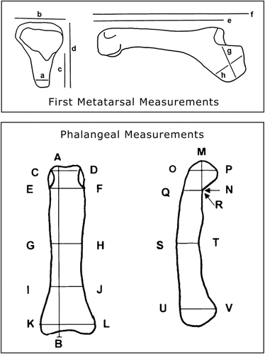
First metatarsal and phalangeal measurements.
RESULTS: DESCRIPTIONS AND COMPARATIVE ANATOMY
Body size
All of the recovered postcranial elements (Table 1) attributed to T. belgica are very small, being similar in size to comparable elements of the smallest living primate, the pygmy mouse lemur, Microcebus berthae (24–38 g; Table 2; see Rasoloarison et al., 2000). Gebo et al. (2012) estimated a conservative size range from 30 to 60 g for T. belgica, and none of these new elements alters this perspective. Recently, Boyer et al. (2013a) have calculated a body weight of 47.25 g for T. belgica, a value well within our size estimation.
| Field ID | Museum | Element | Preservation | Level |
|---|---|---|---|---|
| DIIC2478RS | IRSNB M2163 | Femur | Broken | DIIC |
| DIIC2479RS | IRSNB M2164 | Tibia with fibula | Broken | DIIC |
| DIII2480RS | IRSNB M2165 | Tibia | Broken | DIII |
| DIIC2481RS | IRSNB Vert-32731-01 | Talus | Complete | DIIC |
| DIIC2482RS | IRSNB Vert-32731-02 | Calcaneus | Complete | DIIC |
| DIIC2483RS | IRSNB Vert-32731-03 | Calcaneus | Complete | DIIC |
| DI2484RS | IRSNB Vert-32731-04 | Calcaneus | Complete | DI |
| DIIA2485RS | IRSNB M2166 | First metatarsal | Complete | DIIA |
| DIIC2486RS | IRSNB Vert-32731-05 | First metatarsal | Broken | DIIC |
| DIII2487RS | IRSNB Vert-32731-06 | First metatarsal | Broken | DIII |
| DI2488RS | IRSNB Vert-32731-07 | First metatarsal | Broken | DI |
| DIII2489RS | IRSNB Vert-32731-08 | First metatarsal | Broken | DIII |
| DIIA2490RS | IRSNB M2160 | Humerus | Broken | DIIA |
| DIIA2491RS | IRSNB M2161 | Ulna | Broken | DIIA |
| DIIC2492RS | IRSNB M2162 | Second metacarpal | Complete | DIIC |
| DIIA2493RS | IRSNB M2167 | Manual proximal phalanx | Complete | DIIA |
| DIIC2494RS | IRSNB Vert-32731-09 | Pedal proximal phalanx | Complete | DIIC |
| DIIA2495RS | IRSNB Vert-32731-10 | Proximal phalanx: Digit 1 | Complete | DIIA |
| DI2496RS | IRSNB Vert-32731-11 | Proximal phalanx: Digit 1 | Complete | DI |
| DIII2497RS | IRSNB Vert-32731-12 | Proximal phalanx: Digit 1 | Complete | DIII |
| DIIA2498RS | IRSNB Vert-32731-13 | Pedal proximal phalanx | Complete | DIIA |
| DIII2499RS | IRSNB Vert-32731-14 | Pedal proximal phalanx | Broken | DIII |
| DI2500RS | IRSNB M2168 | Pedal proximal phalanx | Complete | DI |
| DI2501RS | IRSNB Vert-32731-15 | Pedal proximal phalanx | Complete | DI |
| DIIC2502RS | IRSNB Vert-32731-16 | Manual proximal phalanx | Complete | DIIC |
| DIIC2503RS | IRSNB Vert-32731-17 | Pedal proximal phalanx | Broken | DIIC |
| DIII2504RS | IRSNB Vert-32731-18 | Middle phalanx | Complete | DIII |
| DIIC2505RS | IRSNB Vert-32731-19 | Middle phalanx | Complete | DIIC |
| DIIC2506RS | IRSNB Vert-32731-20 | Middle phalanx | Complete | DIIC |
| DIIC2507RS | IRSNB Vert-32731-21 | Middle phalanx | Complete | DIIC |
| DIIC2508RS | IRSNB Vert-32731-22 | Middle phalanx | Complete | DIIC |
- DIRS, DIIARS, DIICRS, and DIIIRS correspond to the four layers mentioned in the work of Smith and Smith (1996).
| Teilhardina belgica | Microcebus berthae (n = 1) | |
|---|---|---|
| Distal humerus width | 4.90 | 4.18 |
| Proximal femur width | 4.90 | 4.75 |
| Distal tibia anterior width | 2.1 | 2.55 |
| Talar length | 4.1–4.35 | 4.47 |
| Calcaneal length | 6.81–7.39 | 8.94 |
| First metatarsal length | 10.35 | 6.32 |
| Lateral-digit proximal phalanx length | 5.15–7.55 | 5.00 |
| Middle phalanx length | 3.25–5.6 | 4.10 |
Distal humerus
The IRSNB M2160 (Royal Belgian Institute of Natural Sciences, Brussels, Belgium) distal left humerus represents the first forelimb element attributed to Teilhardina (Fig. 2). It is broken at the start of the distal shaft and laterally along the brachioradialis flange leaving the elbow joint intact. The comparative measurements to Microcebus berthae are listed in Table 3. In overall elbow anatomy, IRSNB M2160 is very similar to other known omomyids, although smaller with a bicondylar width of only 4.9 mm (Absarokius abbotti and Shoshonus cooperi, two small omomyids, are 6.15 and 6.12 mm in this same dimension, respectively). The round capitulum is slightly taller relative to its width, and this joint exhibits a short capitular tail laterally. The round capitular shape is restricted mediolaterally along the distal edge with a sharp lateral edge similar to Absarokius abbotti (Washakie Basin,?Bitter Creek, UCMP 113301; University of California Museum of Paleontology, Berkeley, CA, USA), Tetonius mckennai (UCMP 134843), Omomys carteri, (UCM 69380 and UCM 69383; University of Colorado Museum, Boulder, CO), and Hemiacodon gracilis (AMNH 29126; American Museum of Natural History, New York, USA); however, it is distinct from the very rounded bottom edge capitular outline of Shoshonius or Tarsius (Dagosto et al., 1999). The T. belgica capitulum is separated from the trochlea by a zona conoidea, a pattern found across primitive primates in both strepsirhine and haplorhine suborders. The trochlea is wide, rather than high, reflecting a more horizontal body posture, in contrast to the tall trochleas found among vertical clinging and leaping primates (Szalay and Dagosto, 1980). Trochlear height to trochlear width ratios (th/tw) confirm this observation across Omomyidae (T. belgica, 0.71; Absarokius, 0.71; Hemiacodon, 0.68–0.74; Omomys, 0.73; Shoshonius, 0.76–0.84; Tetonius, 0.78; microchoerines, 0.68; see Szalay and Dagosto, 1980; Dagosto et al., 1999; Rose et al., 2009). The trochlear width to articular width index (tw/aw) of T. belgica (0.48) is similar to other omomyids (Absarokius, 0.46; Hemiacodon, 0.43–0.48; Omomys, 0.43–0.48; Shoshonius, 0.41–0.47; Tetonius, 0.44; microchoerines, 0.47–0.51; see Szalay and Dagosto, 1980; Dagosto et al., 1999; Rose et al., 2009). The IRSNB M2160 trochlea angles obliquely as in haplorhine primates (Szalay and Dagosto, 1980), and the distal notch between the angle of the distal edge of the trochlea relative to the capitulum is shallow relative to the more sharply v-shaped angular notch in Tarsius, the IVPP V 13022 (Institute of Vertebrate Paleontology and Paleoanthropology, Beijing, China) distal humerus from Shanghuang (China) or Shoshonius. The IRSNB M2160 trochlea more closely resembles taxa such as Absarokius, Tetonius, Omomys, and Hemiacodon. The medial epicondyle of IRSNB M2160 is wide and medially oriented, a characteristic common among primitive primates including other omomyids, whereas the opposite side lateral epicondyle is short. The brachioradialis flange, the attachment site for the brachialis and brachioradialis muscles, is present but quite broken proximally and laterally. The size of all of the muscle attachment sites of this distal humerus implies relatively large arm muscles. IRSNB M2160 has an entepicondylar foramen and a large pit for the humeroulnar ligament posteriorly (i.e., the dorsoepitrochlear fossa), a character that is present among other omomyid and microchoerid distal humeri but is not found in Tarsius (Dagosto et al., 1999). The olecranon fossa is shallow, being wider than high, and the anterior radial fossa is moderate in depth.
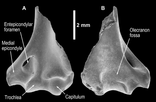
Distal humerus, left; IRSNB M2160; anterior (A) and posterior (B) views.
| T. belgica | M. berthae | |
|---|---|---|
| IRSNB M2160 | EWH 30/147 | |
| Bicondylar width | 4.9 | 4.18 |
| Articular width | 2.9 | 2.95 |
| Capitular width | 1 | 1.09 |
| Capitular height | 1.2 | 1.25 |
| Zona conoidea width | 0.5 | 0.4 |
| Trochlear width | 1.4 | 1.46 |
| Trochlear height | 1 | 1 |
| Medial epicondylar width | 1.5 | 1.6 |
| Lateral epicondylar width | 0.7 | 0.8 |
| Posterior trochlear width | 2 | 1.6 |
| Brachial flange height | (Broken) 3.5 | 6.54 |
| Olecranon fossa height | 1.3 | |
| Olecranon fossa width | 1.75 |
We find no profound differences between the IRSNB M2160 distal humerus and those attributed to other omomyids, for example Absarokius, Hemiacodon, Omomys, Shoshonius, and Tetonius (Szalay and Dagosto, 1980; Dagosto et al., 1999). Relative to Tarsius or to microchoerids such as Microchoerus, IRSNB M2160 differs in a few anatomical features. The trochlea morphology of IRSNB M2160 is neither as tall or as mediolaterally short, as in Tarsius, nor is the capitulum as round or ball-like. The trochlear–capitulum notch is flatter in IRSNB M2160 relative to Tarsius as well. The entepicondylar foramen is smaller and not stretched as obliquely proximally in IRSNB M2160 relative to this foramen in tarsier distal humeri. The radial fossae and medial epicondyles of IRSNB M2160 and Tarsius look very similar to each other. When compared with Microchoerus, IRSNB M2160 has a shorter trochlea and a relatively deeper trochlea–capitular notch (Dagosto, 1993).
Functionally, this elbow reflects a mobile forearm with good radial rotation for pronation and supination of the hand (see Rose, 1993). This elbow joint and muscle attachment anatomy suggests that T. belgica was quite capable of climbing and grasping onto supports reflecting its frequent use of an arboreal habitat.
Proximal ulna
The proximal right ulna (IRSNB M2161) is morphologically different from the more common small ulnae found within the Dormaal collections. The common small ulnae represent rodents (see Emry and Thorington, 1982; Rose and Chinnery, 2004; Thorington et al., 2005), whereas the IRSNB M2161 specimen is rare being the only example of this ulnar morphology in this private collection. IRSNB M2161 differs from the “rodent-like” ulnae in several ways. First, IRSNB M2161, like in other primates, has a more spool-shaped trochlear floor, whereas in rodents, this area is straighter and wider along the medial edge in this region. If the trochlear floor is waisted in rodents, it occurs closer to the proximal end as the trochlear facet rises to the back wall of the trochlear notch in contrast to primates where the waisting occurs distally and in the middle of the joint floor (Conroy, 1976). Second, the olecranon process is medially bent (dorsal view), and the top of the olecranon process is waisted in rodents relative to primates, representing essentially a notch behind the anconeal processes. Third, the brachialis muscle insertion area is less prominent in rodents relative to that of primates and to that of IRSNB M2161. Finally, the anconeal edges at the back of the trochlear notch are straighter and mediolaterally aligned transversely in rodents relative to that of primates (for images of rodent ulnae, see Emry and Thorington, 1982; Rose and Chinnery, 2004; Thorington et al., 2005).
When compared with extant primitive primates, such as galagos or lemurs, or to fossil primates like Notharctus tenebrosus (AMNH 127167) or Cantius trigonodus (USGS 5900; United States Geological Survey, Baltimore, MD), IRSNB M2161 (Fig. 3) compares favorably in terms of proximal joint shape and in two ratio comparisons (Table 4). IRSNB M2161 looks particularly similar to the proximal ulnae of tarsiers. On the basis of these comparisons, we believe that the IRSNB M2161 specimen is best allocated to T. belgica. Unfortunately, the lack of studies establishing solid phylogenetic informative characteristics of the proximal ulna across primate lineages with the exception of Old World monkeys or apes (see Gregory, 1920; Jolly, 1967; Fleagle et al., 1975; Conroy, 1976; Fleagle, 1988; Rose, 1993; Fleagle and Simons, 1995) hampers our ultimate assessment.
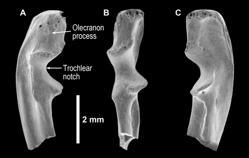
Ulna, right; IRSNB M2161; lateral (A), dorsal (B), and medial (C) views.
| ?primate ulna (T. belgica) | Cantius trigonodus | Notharctus tenebrosus | |
|---|---|---|---|
| IRSNB M2161 | USGS 5900 | AMNH 127167 | |
| Known length | (Broken) 6.35 | (Broken) 34.65 | 89.13 |
| Length of the olecranon notch (lpn) | 2.15 | 6.59 | 8.47 |
| Olecranon process length (opl) | 1.75 | 6.64 | 7.7 |
| Olecranon process width | 1.15 | 5.43 | 7.14 |
| Proximal facet width | 1.4 | 4.75 | 6.04 |
| Minimum facet width | 0.9 | 3.33 | 4.75 |
| Radial facet width (rfw) | 0.75 | 3.39 | 3.72 |
| Total joint width (tjw) | 1.75 | 6.97 | 8.67 |
| Coronoid process width | 1 | 3.34 | 5.72 |
| opl/opl + lpn | 0.45 | 0.50 | 0.48 |
| rfw/tjw | 0.43 | 0.49 | 0.43 |
IRSNB M2161 has a moderately long olecranon process as the attachment site of the triceps muscle. This process extends proximally, not dorsally as in terrestrial quadrupeds (Jolly, 1967; Fleagle et al., 1975; Conroy, 1976; Fleagle, 1988; Fleagle and Simons, 1995), and its length suggests bent elbow body postures with flexion capabilities around 130° (Rose, 1993). The olecranon process is slightly curved medially.
The trochlear or sigmoid notch is deep, being similar in shape to many arboreal primates, including tarsiers. The medial and lateral anconeal processes of the back edge of the trochlear notch curve mediolaterally outward to either side. The proximal joint facet is wide in the middle like many prosimians and New World monkeys, although narrower than either of the proximodistal joint edges, giving this joint a spool-like shape in dorsal view. The anterior edge of the coronoid process has a medially wide joint surface to support the ulna during humero–ulnar flexion. The height of the coronoid process is lower than the height of the olecranon process. The radial notch is aligned along the lateral side of the trochlear notch, being triangular in shape.
The proximal aspect of the ulnar shaft is proximodistally curved with an elongated groove, starting well proximally, along the mid-shaft of the medial side indicating the origin of flexor carpi ulnaris. Just distal to the coronoid process medially is a triangular region for the attachment of flexor digitiorum superficialis, and more distally from this site is the groove for the origin of pronator teres. Above these two muscle sites is a deep groove for the insertion site of a prominent brachialis muscle, a muscle involved in flexion, and a morphological feature similar to that of tarsier ulnae.
The lateral shaft, distal to the radial notch, shows a significant groove for the supinator muscle. Along the ridge that separates the lateral shaft in half lies the site of origin for extensor carpi ulnaris, and below this ridge is where anconeus also attaches.
In terms of ulnar function, the IRSNB M2161 element is clearly not similar to any terrestrial primates or to suspensory apes. It generally matches ulnae from arboreal quadrupedal primates in terms of joint anatomy. IRSNB M2161 shows several deeply grooved surfaces for muscles that flex the elbow or wrist, whereas other bony surfaces support muscles that rotate the radius. In short, like the humerus, the ulna shows a mobile arboreally adapted forelimb with bent elbow positions that suggest a lowering of the body toward a support for better balance (Schmitt, 1999). Again, the forearm of T. belgica was quite capable of using an arboreal habitat.
Metacarpal
Little morphological information exists to determine whether an unassociated metacarpal belongs to a primate (in this case to T. belgica). In contrast to ulnae, however, primate metacarpals are very distinct from those of rodents (Fig. 4). Gregory (1920), Aiello and Dean (1990), Godinot and Beard (1991), Hamrick and Alexander (1996), Lemelin et al. (2008), Franzen (1993), and Boyer et al. (2013b) have all described salient features of primate metacarpals. The bone and joint morphology of IRSNB M2162 compares well with fossil and living primates.
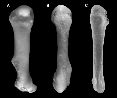
A rodent right second metacarpal (A: Sciurus carolinensis, FMNH 156882) compared with a right second metacarpal of a living primate (B: Otolemur garnettii, NIU 99-7-1), and to IRSNB M2162 (C). All are scaled to the same length.
The IRSNB M2162 second right metacarpal appears relatively short with a large rounded head, being wider than the proximal joint surface (Fig. 5). The metacarpal head joint shape is bulbous and is followed by a sulcus and a slight transverse ridge dorsally. In side view, this joint surface is semicircular. The distal metacarpal head region shows prominent attachment areas for the collateral ligaments. The distal half of this metacarpal shaft is wider or thicker than the proximal half of this bone. The entire shaft exhibits torsion proximodistally from end to end reflecting a supinated hand position. The proximal joint surface is diamond-shaped, being widest at mid-facet. This joint surface is much smaller and flatter than the highly curved distal metacarpal joint surface. Several metacarpal measurements are listed in Table 5.

Second metacarpal, right, IRSNB M2162; distal (A), proximal (B), dorsal (C), plantar (D), lateral (E), and medial (F) views.
| Second metacarpal | Proximal femur | ||
|---|---|---|---|
| IRSNB M2162 | IRSNB M2163 | ||
| Length | 4.9 | Femoral head width | 1.9 |
| Head width | 1.1 | Femoral head height | 1.9 |
| Head height | 1 | Femoral neck length 1 | 2.25 |
| Mid-shaft width | 0.6 | Femoral neck length 2 | 2.5 |
| Mid-shaft height | 0.5 | Proximal width | 4.9 |
| Base width | 0.75 | Mid-shaft ap length | 1.65 |
| Base height | 1 | Mid-shaft ml width | 1.75 |
| Length to third trochanter | 3.6 | ||
| lesser trochanter breadth | 1.1 | ||
| lesser trochanter height | 1.25 | ||
| Third trochanter breadth | 0.5 | ||
| Third trochanter height | 2 | ||
| Trochanteric fossa length | 2.5 |
The comparison of IRSNB M2162 with Notharctus tenebrosus shows no remarkable anatomical differences between the two with the exception of size and robusticity. The IRSNB M2162 metacarpal has a thinner and more elongated shaft, a shallower depression just distal to the proximal joint for carpo-metacarpal ligament, and the proximal joint surface is diamond-shaped rather than quadrate relative to these anterior areas in Notharctus. No remarkable anatomical differences appear when we compare IRSNB M2162 with other living primates such as galagos, lorises, and tarsiers, which generally resemble Notharctus in metacarpal shape. IRSNB M2162 is narrower in the shaft, metacarpal head, and proximal joint surface to these same metacarpal regions in Tarsius. Lemurids and indriids possess relatively longer metacarpals, when compared with other primates and with IRSNB M2162, with lemurs showing broader metacarpal heads relative to indriids. The second metacarpal in primate hands contacts the trapezoid proximally, has a slight dorsal facet with the third metacarpal laterally along with a facet for the capitate, and a slight facet medially with the trapezium or first metacarpal.
In terms of hand function, the broad distal joint surface matches the proximal phalanx joint surface providing a wide contact region for digit support, and the shaft of IRSNB M2162 is elongated for a longer palm region. Overall, these two features support finger flexion for grasping and elongated manual digits in T. belgica.
Proximal femur
The left proximal femur, IRSNB M2163 (Table 5), is broken at the femoral head, the top of the greater trochanter, and at the mid-shaft (Fig. 6). It has a short femoral neck. The head would likely have been about equal to the greater trochanter in height. There is a strong anterior pillar leading to the greater trochanter, and this region of the proximal femur curves anteriorly. The proximal outline of this femur has a square shape if we trace a line from the femoral head to the greater trochanter above to the third and lesser trochanter below, a similar box-like shape pattern to other omomyid femora. In contrast, taxa with a more distally located third trochanter tend to outline a longer or more quadrate proximal femoral outline.
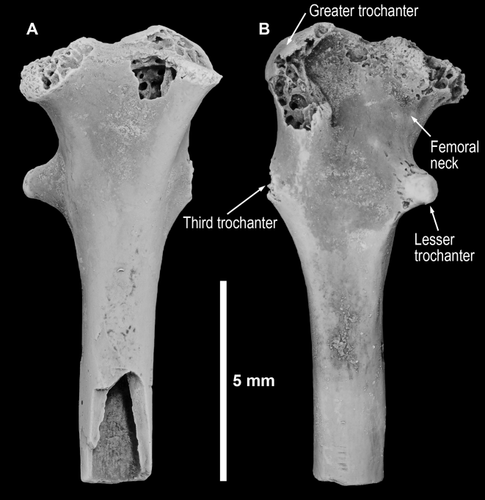
Proximal femur, left; IRSNB M2163; anterior (A) and posterior (B) views.
The greater trochanter is prominent and overhangs the anterior shaft in IRSNB M2163. The lesser trochanter has a slight posterior orientation relative to the midline of the shaft. The third trochanter is oriented directly across from the lesser trochanter at their widest points. The back or posterior aspect of the proximal femur is very flat and wide with a short intertrochanteric fossa, a shared characteristic with other omomyid femora. Lastly, the femoral shaft is mediolaterally wider at the presumed mid-point of the mid-shaft (a–p/m–l = 0.94).
In most aspects, the IRSNB M2163 femur from Dormaal is morphologically very similar to other known femora of omomyids (e.g., Omomys, Hemiacodon, Shoshonius, Ourayia, and Chipetaia; Simpson, 1940; Dagosto and Schmid, 1996; Dagosto et al., 1999; Anemone and Covert, 2000; Dunn et al., 2006; Dunn, 2010). In Shoshonius, the third trochanter is more proximal relative to the lesser trochanter as it is among cheirogaleids, galagos, and tarsiers (Dagosto et al., 1999). In contrast, the third trochanter is directly opposite to the lesser trochanter in IRSNB M2163. The third trochanter is distal to the lesser trochanter among lemurids and indriids and many anthropoids. Anemone and Covert (2000), however, demonstrated variability in trochanter position with all patterns being observed in Omomys.
In terms of functional adaptive abilities, the IRSNB M2163 femur exhibits several features associated with leaping such as an anterior pillar with anteriorly bending of the greater trochanter and a short femoral neck (Dagosto and Schmid, 1996). In contrast to the leaping features, a prominent and medially oriented lesser trochanter and a mediolaterally wider mid-shaft are common features among climbing arboreal primates related to hip flexion (Burr et al., 1982; Dagosto et al., 1999).
Tibiae
Two incomplete right tibiae (IRSNB M2164 with fibula and IRSNB M2165; Fig. 7 and Table 6) have been allocated to T. belgica. IRSNB M2164 has a small piece of the fibula attached to the distolateral side of the tibia, whereas on IRSNB M2165, the fibula is absent. The IRSNB M2164 tibia displays a marked anteroposterior s-shaped curvature as also observed in Shoshonius (Dagosto et al., 1999), both being more curved than in Tarsius. Like Shoshonius and Necrolemur, the IRSNB M2164 tibia has a thin but anteriorly prominent tibial tuberosity (cnemial crest) located far distally from the tibial plateau. On the lateral surface of the shaft adjacent to the crest in is the insertion of the “pes anserinus” (the common tendon of gracilis, sartorius, and semitendinosus; hip and knee flexors). On its medial side is the origin of the tibialis anterior, an ankle dorsiflexor and invertor. The more distal location of the crest is a feature associated with quadrupedalism and differs from the smaller, more proximally located crest of vertical clinging and leaping tarsiers, which may serve to reduce the moment arm for knee flexion during hip extension for leaping primates (Stern, 1971; Fleagle, 1977).
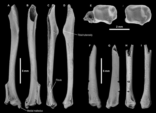
Tibiae, IRSNB M2164 (A–E, right tibia); anterior (A), posterior (B), lateral (C), medial (D), and distal (E) views. IRSNB M2165 (F–J, right tibia); anterior (F), posterior (G), lateral (H), medial (I), and distal (J) views.
| Distal tibia | Distal tibia | |
|---|---|---|
| IRSNB M2164 | IRSNB M2165 | |
| Known length (broken) | 20.48 | 12.74 |
| Mid-shaft anteroposterior length | 1.25 | 1.3 |
| Mid-shaft mediolateral width | 1.5 | 1.4 |
| Length to distal tibia tuberiosity (midpoint) | 5.2 | |
| Anterior width distal tibia | 2.1 | 2.1 |
| Width of tibial malleollus | 1.5 | 1.7 |
| Length of tibial malleollus below the joint | 0.75 | 0.75 |
| Distal joint length | 1 | 1.15 |
| Distal joint width | 1.5 | 1.75 |
| Fibular scar length | 4 | 3.25 |
| Length of fibula | 4.25 | |
| Total distal width (with fibula) | 3.25 |
Both tibiae display a prominent tibiofibular ridge along the distal-most quarter of the tibia. The articulated fibula on IRSNB M2164 and the prominent scar on IRSNB M2165 indicated that although the fibula was unfused to the tibia in T. belgica, it was closely appressed as is also the case in the omomyids Absarokius and Shoshonius, in the fossil anthropoid Apidium, and in marmosets and mouse lemurs (Fleagle and Simons, 1983; Covert and Hamrick, 1993; Dagosto et al., 1999).
In both specimens, the anteroposterior dimension of the moderately grooved talar joint surface slightly exceeds the mediolateral width, as is typically the case in primates. Both tibiae display a marked groove for the tibialis posterior and flexor digitorum longus tendons on the posterior side of the medial malleolus and a wider grooved area laterally for flexor hallucis longus. These grooves are separated by a prominent ridge.
The moderately long tibial malleolus displays a low angle of about 15° relative to the talotibial joint surface, a common condition among haplorhine primates in general and other tarsiiforms in particular (Dagosto, 1985; Dagosto et al., 1999). As the distal end of the fibula is missing, we cannot assess the shape of the tibiofibular mortise. We do, however, know that the fibular facet of tali attributed to T. belgica are straight and steep-sided as in other haplorhine primates, indicating that the mortise was parallel-sided as in other haplorhines rather than asymmetrical as in strepsirhine primates. The tibial malleolus is broad in IRSNB M2164 and IRSNB M2165 tibiae of T. belgica, not reduced posteriorly as in anthropoids (Dagosto et al., 2008).
Tarsals
Tables 1 and 7 provide catalog numbers and measurements for the three new elements; however, given the history of discussion for tarsal elements allocated to T. belgica and our efforts to clarify and figure this tarsal anatomy in Gebo et al. (2012), we see no need to elaborate on comments since our 2012 publication. We do note that the three new calcanei length measures (6.9, 7.2, and 7.3 mm) fall in the upper range of previous calcaneal length measures and that the IRSNB Vert-32731-02 calcaneus at 7.3 mm in total length is similar to the relatively larger specimen (IRSNB M1236; 7.39 mm) discussed in Gebo et al. (2012). Overall, the nine calcanei that we have been able to measure from Dormaal demonstrate a length range from 6.52 to 7.39 mm, a range that spans less than 1 mm from the lowest to the highest value with incremental increases in length across these nine specimens (6.52, 6.62, 6.83, 6.9, 6.98, 7.2, 7.21, 7.3, and 7.39 mm), supporting our hypothesis that the sample represents a single species of T. belgica at Dormaal.
| Talus | IRSNB Vert-32731-01 | Calcaneus | IRSNB Vert-32731-02 | IRSNB Vert-32731-03 | IRSNB Vert-32731-04 |
|---|---|---|---|---|---|
| Talar length | 4.35 | Calcaneal length | 7.3 | 6.9 | 7.2 |
| Talar width | 2.35 | Distal length | 4.15 | 3.7 | 3.65 |
| Trochlea length | 1.7 | Posterior calcaneal facet width length | 1.5 | 1.5 | 1.85 |
| Mid-trochlear width | 1.75 | Posterior calcaneal facet width | 0.6 | 0.8 | 0.8 |
| Talar head height | 1.25 | Heel length | 1.85 | 1.65 | 1.6 |
| Talar head width | 1.55 | Calcaneocuboid facet width | 1.3 | 1.35 | 1.8 |
| Lateral body height | 2 | Calcaneocuboid facet height | 1.25 | 1.15 | 1.3 |
| Lateral body length | 1.55 | Calcaneal width | 2.15 | 2.35 | 2.65 |
| Posterior calcaneal facet maximum width | 0.9 | ||||
| Posterior calcaneal facet minimum width | 0.6 | ||||
| Posterior calcaneal facet length | 1.6 | ||||
| Talar neck length | 2.5 |
First metatarsals
The first metatarsal is one of the most distinctive elements of the primate postcranium and cannot be mistaken for any other (Szalay and Dagosto, 1988). In 2012, we addressed the anatomy of the proximal joint surface in T. belgica, and now we can add a description and comparison of the complete element. We recovered five additional first metatarsals. Four preserve only the proximal end but one is almost completely preserved (IRSNB M2166), missing only the dorsal aspect of the proximal joint region (Fig. 8). It is surprisingly long at 10.35 mm given Teilhardina's overall size similarity to M. berthae whose first metatarsal measures only 6.32 mm (or 61% of the length of IRSNB M2166. A long first metatarsal may be another manifestation of a long foot, as suggested by the long phalanges documented in Gebo et al. (2012) and below.
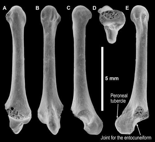
First metatarsal (right), IRSNB M2166; dorsal (A), plantar (B), lateral (C), proximal (D), and medial (E) views.
In Gebo et al. (2012), we described the proximal joint anatomy noting the prominent and long peroneal tubercle. The more complete length of the IRSNB M2166 specimen allows us to comment on the shaft as well as to construct several ratios (see Table 8). The shaft in IRSNB M2166 is relatively narrow from mid-shaft distally and fairly straight along the plantar edge. This shape compares well with a small primate like Microcebus or to the haplorhine Tarsius but is unlike with the first metatarsal shape in adapiforms or lemuriforms, which are more robust and have greater curvature. The IRSNB M2166 shaft of Teilhardina is similar in shape to that of Arapahovius (figured in Szalay and Dagosto, 1988).
| IRSNB M2162 | IRSNB M2166 | IRSNB Vert-32731-05 | IRSNB Vert-32731-06 | IRSNB Vert-32731-07 | IRSNB Vert-32731-08 | Mean | |
|---|---|---|---|---|---|---|---|
| Peroneal tubercle width, a | 0.70 | 0.45 | 0.55 | 0.6 | 0.58 | ||
| Proximal width, b | 2.15 | 2.15 | 2.15 | 2 | 2.11 | ||
| Peroneal tubercle length, c | 1.25 | 1.5 | 1.35 | 1.5 | 1.40 | ||
| Dorsoplanar depth of proximal metatarsal, d | 2.40 | 2.75 | 2.3 | 2.5 | 2.49 | ||
| Dorsal length of shaft and head, e | 9.2 | 9.2 | |||||
| Total length, f | 10.35 | 10.35 | |||||
| Lateral tubercle length, g | 1.70 | 1.5 | 1.5 | 1.45 | 1.54 | ||
| Lateral tubercle height, h | 1.50 | 1.25 | 1.25 | 1.35 | 1.34 | ||
| Head width | 1.8 | 1.5 | 1.5 | 1.60 | |||
| Head height | 1.5 | 1.15 | 1.4 | 1.35 | |||
| Mid-shaft width | 0.9 | 0.85 | 0.85 | 0.9 | 0.88 | ||
| Mid-shaft height | 0.9 | 0.85 | 0.85 | 1 | 0.90 | ||
| Ratios | |||||||
| a/b | 0.33 | 0.21 | 0.26 | 0.30 | 0.28 | ||
| c/d | 0.52 | 0.55 | 0.59 | 0.60 | 0.57 | ||
| e/f | 0.89 | 0.89 | |||||
| h/g | 0.88 | 0.83 | 0.83 | 0.93 | 0.87 | ||
| d–c/b | 0.53 | 0.58 | 0.44 | 0.50 | 0.52 |
The head of the IRSNB M2166 first right metatarsal shows the primate asymmetrical arrangement of the tubercles with a bulbous distal joint surface extending outward in the middle. The plantar (medial) tubercle of the head extends more prominently outward than the dorsal (lateral) tubercle, and the plantar tubercle is shorter proximodistally when compared with the dorsal tubercle. The head anatomy appears similar to many different primate groups and is not distinctive by itself.
Comparative proportions of first metarsal measurements are given in Table 8. T. belgica is similar to other omomyids (Shoshonius, Omomys, and Hemiacodon in Gebo et al., 2012) in the a/b ratio, with values that indicate a narrow tubercle. The c/d ratio compares tubercle length with the length of the proximal end, and the lower value of T. belgica indicates a relatively shorter tubercle, a similarity with other primitive Eocene primates. The c/d ratio for T. belgica, however, is the lowest among omomyids. The e/f ratio, which compares shaft length to total length, is 0.89, the same as Hemiacodon and to Omomys (0.83) and Necrolemur (0.88). The h/g ratio (0.87), which compares height and length of the peroneal tubercle, is most similar to Omomys (0.89). The Omomys and Teilhardina ratio values are lower relative to other known omomyids and microchoerids ratios with values above 0.94. The d–c/b ratio compares the height of the proximal joint surface relative to the width of the first metatarsal base. T. belgica (0.52) is similar to Shoshonius (0.49), Hemiacodon (0.49), Omomys (0.59), and Necrolemur (0.57; Gebo et al., 2012). As indicated by these ratios, T. belgica exhibits similarities to other omomyid genera in first metatarsal shape, suggesting shared proportionality among these related taxa.
First-digit proximal phalanges
Two proximal first-digit phalanges were discussed in Gebo et al. (2012). Three additional first-digit proximal phalanges have been recovered here (IRSNB Vert-32731-10, IRSNB Vert-32731-11, and IRSNB Vert-32731-12; Table 9). The three new specimens are slightly shorter (4.15–4.35 mm in length) than the two previously described. IRSNB Vert-32731-11 is a juvenile with an epiphyseal line present. All three of the new elements are robust and appear anatomically most similar to the IRSNB 1265 element, which was allocated to the foot by Gebo et al. (2012) on the basis of the anatomy of the proximal base. We allocate these specimens to the foot as well. Table 9 shows that the ratio values of the three specimens are very similar to each other. We note again that the IRSNB 1264 specimen allocated to the hand is relatively longer (AB/KL phalangeal length to base width ratio, 3.52) than IRSNB 1265 (2.97), IRSNB Vert-32731-10 (2.90), IRSNB Vert-32731-11 (2.77), or IRSNB Vert-32731-12 (2.8). This is a similarity to tarsier manual first digits (Gebo et al., 2012). The T. belgica proximal pedal phalangeal ratios for AB/KL are also long relative to basal width as in tarsiers (2.87), but much longer than in galagos (2.2-2.3; Gebo et al., 2012). Elongation is the one notable distinction of the T. belgica first-digit proximal elements, especially for the manual element.
| IRSNB Vert-32731-10 | IRSNB Vert-32731-11 | IRSNB Vert-32731-12 | Mean | |
|---|---|---|---|---|
| AB | 4.35 | 4.15 | 4.25 | 4.25 |
| CD | 0.75 | 0.75 | 0.75 | |
| EF | 1.25 | 1.25 | 1.25 | |
| GH | 0.8 | 0.75 | 0.8 | 0.78 |
| IJ | 1 | 1 | 1 | 1.00 |
| KL | 1.5 | 1.5 | 1.5 | 1.50 |
| MN | 0.7 | 0.75 | 0.75 | 0.73 |
| OP | 0.6 | 0.7 | 0.75 | 0.68 |
| QR | 0.45 | 0.5 | 0.5 | 0.48 |
| ST | 0.65 | 0.7 | 0.65 | 0.67 |
| UV | 1.15 | 1.2 | 1.05 | 1.13 |
Lateral-digit proximal phalanges
Eight lateral-digit proximal phalanges that compare well morphologically with living and extinct primates (see Gregory, 1920; Franzen, 1993; Hamrick et al., 1995) have been identified from Dormaal (Figs. 9 and 10 and Table 10). Six specimens are provisionally allocated to lateral proximal pedal phalanges (IRSNB M2168, IRSNB Vert-32731-09, IRSNB Vert-32731-13, IRSNB Vert-32731-14, IRSNB Vert-32731-15, and IRSNB Vert-32731-17), and two specimens are allocated as proximal manual elements (IRSNB M2167 and IRSNB Vert-32731-16). IRSNB Vert-32731-14 and IRSNB Vert-32731-17 are broken. The longest specimens are allocated to the foot, and the measurable elements are IRSNB Vert-32731-13 (7.55 mm), IRSNB Vert-32731-15 (5.7 mm), and IRSNB M2168 (5.25 mm). IRSNB Vert-32731-13 is very long relative to the other two measurable specimens. The manual lateral phalanges measure 5.15 and 5.6 mm in length. In terms of the length to proximal width ratio (AB/KL), all pedal (5.18–5.83) and manual (5.15–5.6) elements show high ratio values, a similarity to the same ratio values calculated for the middle phalanges (Gebo et al., 2012). This implies long lateral digits (2–5) in the hands and feet of T. belgica.
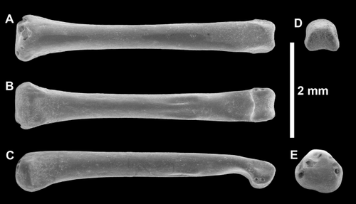
Manual proximal phalanx, IRSNB M2167; dorsal (A), plantar (B), lateral (C), distal (D), and proximal (E) views.
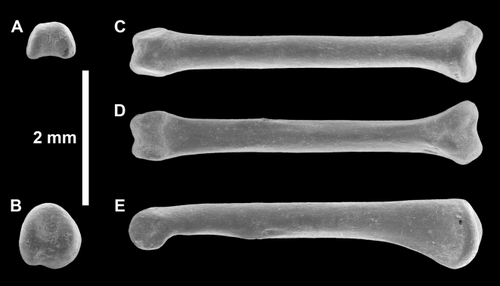
Pedal proximal phalanx, IRSNB M2168; distal (A), proximal (B), dorsal (C), plantar (D), and lateral (E) views.
| Pedal | |||||||
|---|---|---|---|---|---|---|---|
| IRSNB Vert-32731-13 | IRSNB Vert-32731-14 | IRSNB M2168 | IRSNB Vert-32731-15 | IRSNB Vert-32731-17 | IRSNB Vert-32731-09 | Mean | |
| AB | 7.55 | 5.25 | 5.7 | 5.35 | 5.96 | ||
| CD | 0.6 | 0.5 | 0.5 | 0.45 | 0.5 | 0.4 | 0.49 |
| EF | 1 | 0.75 | 0.75 | 0.7 | 0.85 | 0.7 | 0.79 |
| GH | 0.6 | 0.5 | 0.5 | 0.4 | 0.65 | 0.5 | 0.53 |
| IJ | 0.8 | 0.7 | 0.6 | 0.7 | 0.75 | 0.7 | 0.71 |
| KL | 1.3 | 0.9 | 1.1 | 1 | 1.08 | ||
| MN | 0.7 | 0.65 | 0.6 | 0.65 | 0.7 | 0.55 | 0.64 |
| OP | 0.8 | 0.6 | 0.55 | 0.5 | 0.65 | 0.5 | 0.60 |
| QR | 0.5 | 0.4 | 0.45 | 0.35 | 0.4 | 0.35 | 0.41 |
| ST | 0.65 | 0.5 | 0.55 | 0.5 | 0.55 | 0.5 | 0.54 |
| UV | 1.35 | 1 | 1.1 | 0.9 | 1.09 | ||
| Manual | Omomys carteri, manual | ||||||
| IRSNB M2167 | IRSNB Vert-32731-16 | Mean | UCM 69339 | ||||
| AB | 5.6 | 5.15 | 5.38 | 12.22 | |||
| CD | 0.5 | 0.4 | 0.45 | 1.66 | |||
| EF | 0.7 | 0.75 | 0.73 | 2 | |||
| GH | 0.5 | 0.45 | 0.48 | 1.44 | |||
| IJ | 0.75 | 0.65 | 0.70 | 1.89 | |||
| KL | 1 | 1 | 1.00 | 2.55 | |||
| MN | 0.6 | 0.6 | 0.60 | 1.44 | |||
| OP | 0.6 | 0.6 | 0.60 | 1.55 | |||
| QR | 0.35 | 0.45 | 0.40 | 0.89 | |||
| ST | 0.5 | 0.5 | 0.50 | 1.11 | |||
| UV | 0.85 | 1 | 0.93 | 1.89 | |||
These lateral proximal phalanges are very straight along the plantar edge of the shaft, a similarity with tarsier phalanges. Most other primates display more curvature along this edge. Boyer et al. (2013b; Table 9) noted angles of phalangeal curvature for a variety of living and fossil primates. In Boyer et al. (2013b), tarsiers, Apidium, an unspecified omomyid, and Archaeolemur show the lowest mean values for curvature. The manual phalanges of T. belgica narrow at mid-shaft and show slight ridging for the flexor sheaths with a wide hood-like distal shaft. The shaft is very straight, whereas the distal head and joint surface angles plantarly. Proximal to the head is a thinned neck region leading to the distal head and joint. The distal head shows medial and lateral depressions for the collateral ligaments in both manual and pedal phalanges. The proximal manual phalangeal joint is transversely straight and generally a flattened joint surface. In contrast, the pedal phalanges differ in being basally taller proximally with an extensor depression dorsally and a notched joint surface proximally, reflecting the narrower metatarsal head shapes relative to that of metacarpals. The shaft is straighter among these pedal phalanges without any mid-shaft narrowing, and the flexor sheaths are slightly more prominent. The neck and distal head region in the pedal phalanges lacks the great narrowing found in the manual lateral phalanges. Otherwise, the manual and pedal lateral proximal phalanges are very similar morphologically.
Middle phalanges
Two middle phalanges (IRSNB Vert 26857-04 and IRSNB M1266) were allocated to T. belgica by Gebo et al. (2012). Here, we add five additional middle phalanges to the known sample (Table 11). Three of the new specimens are similar in length (4.6–5.2 mm) to IRSNB M1266; however, two IRSNB Vert-32731-20 and IRSNB Vert-32731-19 are significantly shorter (3.25 and 3.35 mm).
| Manual | Pedal | ||||||
|---|---|---|---|---|---|---|---|
| IRSNB Vert-32731-18 | IRSNB Vert-32731-21 | IRSNB Vert-32731-22 | Mean | IRSNB Vert-32731-19 | IRSNB Vert-32731-20 | Mean | |
| AB | 4.6 | 4.35 | 5.2 | 4.72 | 3.35 | 3.25 | 3.30 |
| CD | 0.5 | 0.4 | 0.5 | 0.47 | 0.5 | 0.45 | 0.48 |
| EF | 0.75 | 0.65 | 0.75 | 0.72 | 0.6 | 0.65 | 0.63 |
| GH | 0.65 | 0.65 | 0.6 | 0.63 | 0.5 | 0.35 | 0.43 |
| IJ | 0.75 | 0.75 | 0.8 | 0.77 | 0.7 | 0.6 | 0.65 |
| KL | 1.05 | 1 | 1.1 | 1.05 | 0.95 | 0.9 | 0.93 |
| MN | 0.35 | 0.45 | 0.5 | 0.43 | 0.35 | 0.4 | 0.38 |
| OP | 0.45 | 0.4 | 0.45 | 0.43 | 0.35 | 0.4 | 0.38 |
| QR | 0.25 | 0.3 | 0.35 | 0.30 | 0.25 | 0.3 | 0.28 |
| ST | 0.5 | 0.45 | 0.5 | 0.48 | 0.5 | 0.45 | 0.48 |
| UV | 0.85 | 0.85 | 0.95 | 0.88 | 0.75 | 0.8 | 0.78 |
Although we noted in 2012 and repeat now the difficulty in identifying anatomical features that consistently distinguish manual from pedal middle phalanges, making any definitive allocation to the hand or foot very difficult across primates and for T. belgica, we thought the great elongation of these middle phalanges as highly unusual among primates, especially at this tiny size range. The only living primate where a similar anatomical pattern occurs is in tarsier hands where we find manual middle phalanges being quite long relative to their respective pedal elements. The most complete specimen, IRSNB M1266 described in Gebo et al. (2012), is in fact very similar in length to the much larger Tarsius syrichta, suggesting that these long middle phalanges might possibly represent manual elements within the hands of T. belgica, as is the case in tarsier hands relative to their feet.
There are two ways to interpret this size distinction. One view is to suggest that the different sized middle phalanges are due to the five different digit lengths in both the hand and the foot. A second approach, advocated here, is to place the longer middle phalanges in the hand, like what we observe in tarsiers, and the shorter specimens within the foot of T. belgica. This view accounts for the two apparent size classes of the six measureable middle phalangeal specimens, a small sample size no matter the interpretation. If this second view is correct, Teilhardina is noted to possess very long fingers as in living tarsiers and should use them in similar functional and behavioral ways (see Gebo et al., 2012; Boyer et al., 2013b). In terms of overall anatomy, we have nothing to add to our initial descriptions or comparisons of primate middle phalanges made in Gebo et al. (2012).
Hand and foot proportions
Lemelin (1999) created several digital indices to analyze hand and foot digital rays by adding proximal and middle phalangeal lengths and then dividing by their respective metacarpal ray length. Only the second metacarpal is known in T. belgica, thus our comparisons are limited to that digit. In T. belgica, the ratio of phalangeal length to metacarpal length (calculated on the basis of mean phalangeal lengths) is 210.6, a value that exceeds that of cheirogaleids. For Ray 2, a small mouse-sized lemur like Microcebus murinus has a mean ratio value of 142.2, indicating long phalanges relative to its second metacarpal (Lemelin, 1999). In the same way, Cheirogaeus medius (136.1) and Cheirogaleus major (131.6) also display similarly high ratio values. The highest mean ratio values from Lemelin (1999) are from Digit 4 among these three extant taxa with values from 164 to 182.5. If we use the lowest phalangeal lengths for Teilhardina, this ratio calculation [(5.15 + 4.35)/5.38] is 176.6, a value still high for cheirogaleid second digits but a value within the range of their longest fourth digits.
Hamrick (2001) also analyzed manual ray proportions by calculating digital ray length by combining lengths of the metacarpal, proximal phalanx, and middle phalanx for the third digit. As T. belgica only has a second metacarpal, we substituted this length as the next best comparison with extant species analyzed by Hamrick (2001). The T. belgica ratio for relative metacarpal length to the length of the digital ray (MCL) is exactly the same as that of a tarsier (MCL% = 32.2), a value lower than the other extant primates in the Hamrick (2001) sample, and a ratio value implying a relatively shorter palm to finger lengths. Most primates listed in Hamrick (2001) show values in the mid to high thirties or above for this ratio.
The proximal phalanx to digital ray length for T. belgica (PPL% = 35.3) is proportionally lower than tarsier or loris ratios but is similar to the ratios calculated for platyrrhines and cheirogaleids (Hamrick, 2001). The intermediate or middle phalangeal length relative to digit ray length for T. belgica is longer (IPL% = 32.5) than the mean values reported by Hamrick (2001), including tarsiers (28.4), who possess the highest mean ratio for this comparison. This implies a very long middle phalangeal segment for T. belgica. Lastly, both the proximal and middle phalangeal lengths were compared with the length of the metacarpal in Hamrick (2001), and this ratio (PPL/MCL) is 109.7 for T. belgica, a similar value relative to Loris (109.2) and Galago (107.6) but a value lower than Tarsius (122.1; Hamrick, 2001). Hamrick (2001) noted that primates differ from archontan mammals in possessing a short metacarpal and long proximal phalange as does T. belgica. Proximal phalanx to metacarpal lengths for Adapis, Europolemur, and Notharctus are above 100 for this ratio comparison (Hamrick, 2001) as it is for T. belgica (110).
These length comparisons show how T. belgica divides its hand segments into approximate thirds (metacarpal: 32.2%; proximal phalanx: 35.3%; and middle phalanx: 32.5%), with the longest segment represented by the proximal phalanx. Overall, the metacarpal segment within the hand of T. belgica is the same in relative proportional length to tarsier hands, whereas the middle phalanx is even longer than the noted elongated middle phalanges of tarsier hands. However, the longest digital ray segment belongs to the proximal phalanges of T. belgica. Tarsiers hands show greater elongation in the proximal phalanx (39.2%) region relative to the other segments of their digital ray (32.2%, 39.2%, and 28.4%; Hamrick, 2001).
A relatively shorter palm with long fingers, especially the proximal and middle phalanges, characterizes T. belgica. The long-fingered hands of tarsiers are known as a trait that can effectively grab slender branches as well as small insects (Napier, 1961; Niemitz, 1984; Lemelin, 1999; Hamrick, 2001). With its long manual digits, especially relative to its body size, T. belgica may have used a fast two-handed digital capture technique for active insects that maximized hand spatial area as a net as noted for tarsiers (Lemelin and Jungers, 2007).
Lemelin (1999) also calculated a similar segmental ratio comparison for the hallux (proximal phalanx length divided by first metatarsal length). The T. belgica value for this hallucal ratio is 41.5, a low value relative to Microcebus murinus (57.8), Cheirogaleus medius (52.9), and Cheirogaleus major (55.2; Lemelin, 1999). A lower ratio value implies a long first metatarsal relative to the proximal phalanx, and all of the cheirogaleids show values about half of the length of the first metatarsal. In terms of relative hallucal proportions, T. belgica is even longer, given its lower ratio value, suggesting that the hallucal first digit of T. belgica was particularly long.
Recently, Boyer et al. (2013b) compared hands and digits across a variety of early fossil primates, extant primates, and plesiadapiforms. They documented many interesting comparisons, given the known fossil material that is available, mostly adapiforms or plesiadapiforms. They noted one similarity to T. belgica in that a long middle to proximal phalangeal ratio occurs for the adapiform, Godinotia neglecta, as well. Unfortunately, no omomyid hands exist that would allow a detailed comparison with our few phalanges of Teilhardina.
In a similar manner, Kirk et al. (2008) examined hand proportions across euarchontans and other mammals and noted with Lemelin (1999) and Hamrick (2001) that living and Eocene fossil primates demonstrate an elongation of their manual digits attributed to long proximal phalanges relative to metacarpal length, a feature also noted for T. belgica.
DISCUSSION
Adaptation
The new postcranial elements reported here and the ones described in Gebo et al. (2012) for T. belgica allow a partial reconstruction of the skeleton of one of the most primitive haplorhine primates as well as an assessment of its positional behavioral capabilities. The forelimb of Teilhardina indicates an arboreal primate with good elbow rotational mobility, given the roundness of the capitulum and the spatial separation via the zona conoidea from the downturned trochlea. The trochlea is mediolaterally wide, rather than tall, reflecting a horizontal, rather than a vertical body posture (see Szalay and Dagosto, 1980). The downturned or obliquely angled trochlea, a known haplorhine character (Szalay and Dagosto, 1980), implies that the ulna is more laterally oriented toward the wrist relative to the elbow, making the forearm more widely splayed relative to a more anteroposterior straight-line orientation from the trochlea to the wrist. This laterally deviated forearm spreads the hands away from the midline of the body. The angle of incline suggests that the hands of haplorhines, including those of Teilhardina, are often further away from a mid-line body position with the forelimb held in a vertical or semisupinated hand position with the ulna positioned below the radius. These lateral hand positions imply grasps that are spread further apart and are adaptive for relatively large diameter supports given the small body sizes, often below 100 g for early haplorhines like Teilhardina. A similar functional forelimb argument has independently been made for Plesiadapis cookei (see Boyer, 2009).
The distal humerus of T. belgica is wide with prominent medial and lateral epicondyles suggesting muscular forearm flexors and extensors. The brachioradialis flange and the prominent groove for the brachialis insertion are other indications of prominent forearm musculature. The proximal ulna has several grooves and depressions also suggestive of prominent forelimb muscles (i.e., flexor carpi ulnaris, flexor digitiorum superficialis, pronator teres, supinator, extensor carpi ulnaris, and anconeus). All imply forceful forelimb-grasping capabilities. The moderately long olecranon process implies bent elbow body postures with flexion capabilities around 130° (Rose, 1993). The olecranon fossa of the humerus is shallow, and this also reflects a flexed elbow posture.
The hand of Teilhardina is long with very long fingers, especially the middle phalanges. A relatively short palm and long fingers characterize T. belgica as this taxon displays a relatively shorter metacarpal relative to its longer proximal phalanges, a known characteristic of primate hands (Hamrick, 2001; Kirk et al., 2008; Boyer et al., 2013b). The hand segments of T. belgica are distributed into approximate thirds, with the proximal phalangeal segment being the longest. The long-fingered hands of tarsiers act as a spatial net to digitally capture active insects (Napier, 1961; Lemelin, 1999; Hamrick, 2001; Lemelin and Jungers, 2007), and this may have been the case for T. belgica as well.
T. belgica has several hindlimb characteristics associated with frequent leaping, arboreal quadrupedalism, climbing, and grasping. The proximal femur has a short and straight femoral neck with a femoral head about equal in height to the greater trochanter. There is a strong anterior pillar on the anterior surface of the proximal femur, and this region curves anteroposteriorly with an overhanging greater trochanter. The third trochanter is proximally positioned. The knee is characterized by a tall patellar joint with an elevated lateral patellar rim. All of these femoral features are similar to other known omomyid femora and have been interpreted to indicate leaping specializations (Napier and Walker, 1967; McArdle, 1981; Anemone, 1990; Dagosto, 1993; Dagosto and Schmid, 1996; Dagosto et al., 1999; Anemone and Covert, 2000; Dunn et al., 2006; Dunn, 2010). The two distal tibiae attributed to T. belgica are long and s-curved with a closely apposed fibula, features associated with frequent leaping primates as well (Fleagle and Simons, 1983; Covert and Hamrick, 1993; Dagosto et al., 1999). In the foot, the tarsal anatomy of T. belgica with its distally elongated and narrow calcanei and long talar necks reflect specializations for leaping (Gebo et al., 2012).
The prominent and medially oriented lesser trochanter and a mediolaterally wider femoral shaft imply hip flexion and climbing abilities (Burr et al., 1982; Dagosto et al., 1999). The distal location of the tibial crest is associated with arboreal quadrupedalism relative to vertical clinging and leaping primates (Stern, 1971; Fleagle, 1977). The distal tibiae show a marked depression for toe flexor tendons or muscle tendons associated with inverting the foot, adaptive features related to grasping and climbing. The tibial–fibular mortise appears to be parallel sided via the bony protuberances from the tibial and fibular malleoli demonstrating a joint adapted for fore and aft movements at the upper ankle joint (Dagosto, 1985).
The shape of the articulation of the transverse tarsal and lower ankle joints imply good inversion capabilities while grasping curved substrates in T. belgica. The long first metatarsal of T. belgica (1.6 times as long as that of Microcebus berthae) as well as its proximal joint anatomy noted previously (Gebo et al., 2012) are both highly indicative of a well-developed grasping and an opposable big toe, as is the robust proximal phalanx anatomy. The long and well-muscled hallux of T. belgica can be coupled with long lateral phalanges to reconstruct a foot with long grasping digits. Long toes facilitate the grasping of relatively larger curved substrates in a small primate with the size of T. belgica. The demonstration of a flat hallucal nail in T. brandti (Rose et al., 2011) complements this grasping assessment as no distal phalanges are known for T. belgica.
The phylogenetic position of Teilhardina
In a recent analysis including postcranial characters, Ni et al. (2013) established the position of T. belgica as a relatively primitive tarsiiform primate. None of the new materials presented here would alter that assessment. In fact, many features of the distal humerus, proximal ulna, and distal tibia recognized as primate synapomorphies in Ni et al. (2013) are now known to be present in Teilhardina. Similarly, in addition to tarsal features typical of haplorhines, T. belgica now exhibits haplorhine synapomorphies of tibial malleolar shape and humeral trochlear shape. Its placement among Tarsiiformes was supported previously by several characters of the tarsal bones and is now buttressed by olecranon fossa depth, reduced size of the third trochanter of the femur, and the strong curvature of the tibial shaft. It is more difficult to assess Teilhardina's position within Tarsiiformes because the lack of knowledge of the postcrania of the other taxa and the basal position suggested by Ni et al. (2013) is based primarily on dental attributes.
Many skeletal elements (e.g., the elbow, hip, tibia, and tarsals) attributed to T. belgica are morphologically quite similar to known elements of other omomyids. The ulna, however, attributed to T. belgica is morphologically similar to that of a living tarsier; however, this bone is unknown among other omomyids. The second metacarpal, as well as finger and toe bones, are also unknown in other omomyids, with the exception of a few phalanges attributed to Omomys and to probably Anemorhysis (see Boyer et al., 2013b), and therefore, we cannot adequately compare Teilhardina with most omomyid taxa.
With these limitations in mind and given the primitive phyletic status of Teilhardina as well as the new early Eocene fossils described from Hubei, China (Ni et al., 2013), and Vastan, India (Rose et al., 2009), we believe it is worth reassessing the ancestral skeletal anatomy for Order Primates and for the origin of Tarsiiformes (Table 12). For example, humeral anatomy is consistently similar in morphology across Teilhardina, other omomyids such as Shoshonius, tarsiers, Archicebus, asiadapines, notharctines, and cercamoniines. All display a round humeral head that is situated slightly above the greater tubercle where known as well as a round capitulum, a zona conoidea, and a wide trochlea at the elbow joint (Gregory, 1920; Szalay and Dagosto, 1980; Dagosto et al., 1999; Rose et al., 2009; Ni et al., 2013). The commonality of these features across these groups suggests ancestral primate status for these morphologies. These early primates differ, however, in where the deltopectoral crest is located. For example, Shoshonius displays a more proximally located deltopectoral crest (Dagosto et al., 1999), whereas this crest is distal in asiadapines (Rose et al., 2009). Other morphological distinctions between primate groups exist concerning the size and length of the brachioradialis flange or in the angle of the trochlea (Szalay and Dagosto, 1980). The brachioradialis flange is prominent in the distal humeri of omomyids and in notharctines (Szalay and Dagosto, 1980) but less so among asiadapines, being smaller and less projected laterally. The trochlea is downturned medially among haplorhines, whereas this joint edge is horizontal in its orientation in strepsirhines like the adapiforms (Szalay and Dagosto, 1980), including the asiadapines (Rose et al., 2009). The type of features that differ between early primate clades require outgroup assessments for an ancestral primate characterization, given their lack of uniformity, whereas all early Eocene fossil primates possess ulnae with a long olecranon process, a more uniform feature.
| Character | Anatomical character | Teilhardina belgica (+ other omomyids) | Archicebus | Asiadapines (Marcgodinotius and Asiadapis) | Cantius (+ Notharctus) |
|---|---|---|---|---|---|
| 1 | Humeral head location | ? (head above tubercle in Shoshonius) | Head above tubercle | Head above tubercle | Head above tubercle |
| 2 | Position of deltoid tuberosity | ? (proximal in Shoshonius) | ? | More distal than Shoshonius | More distal than Shoshonius |
| 3 | Brachioradialis flange | Present (tall and wide in Shoshonius) | Present | Tall and narrow | Tall and wide |
| 4 | Trochlear angle | Oblique | Oblique | Horizontal | Horizontal |
| 5 | Capitular shape | Round | Round | Round | Round |
| 6 | Zona conoidea | Present | Present | Present | Present |
| 7 | Metacarpal length | Moderate | ? | ? | Short |
| 8 | Manual phalanges | Long | ? (toes are long) | ? | Long |
| 9 | Shape of ilium | ? (rod-like in Omomys) | Rod-like | ? | Wide ilium |
| 10 | Ischium length | ? (long in Omomys) | Long | ? | Long |
| 11 | Femoral head shape | ? (semicylcindrical in other omomyids) | Semicylindrical | Obliquely round | Obliquely round |
| 12 | Femoral neck length | Short | Short | Moderately long | Moderately long |
| 13 | Third trochanter position | Proximal | Proximal | Distal | Distal |
| 14 | Angle of lesser trochanter | Medial, slightly posteriorly | Medial, slightly posteriorly | Medial, slightly posteriorly | Medial, slightly posteriorly |
| 15 | Height of distal femur | Tall | Tall | Moderate | Tall |
| 16 | Lateral patellar rim | Highest rim | Highest rim | Equal to medial rim | Highest rim |
| 17 | Patellar groove width | Narrow | Narrow | Narrow | Narrow |
| 18 | Tibial plateau spines | ? (2, Hemiacodon; 1, Ourayia) | 2 | ? | 1 |
| 19 | Length of tibia | Long | Long | ? | Moderate |
| 20 | Position of distal fibular | Closely apposed | Closely apposed | ? | Separate |
| 21 | Angle of tibial malleolus | Straight or less angled | Straight or less angled | ? | Oblique |
| 22 | Talofibular mortise | Steep-sided | Steep-sided | ? | Oblique laterally |
| 23 | Talar neck length | Long | ? | Moderately long | Moderately long |
| 24 | Medial talotibial facet | Full | Full | Full | Full |
| 25 | Talofibular facet | Steep | Steep | Oblique | Oblique |
| 26 | Posterior trochlear shelf | Small | ? | Well-developed | Well-developed |
| 27 | Location of flexor hallucis longus groove | Mid-line except perhaps for T. brandti | ? | Lateral | Lateral |
| 28 | Calcaneal shape | Narrow | Wide | Wide | Wide |
| 29 | Distal calcaneal length | Elongated | Elongated | Short | Short |
| 30 | Calcaneocuboid joint shape | Fan-shaped, central pivot | Fan-shaped, central pivot | Fan-shaped, central pivot | Fan-shaped, central pivot |
| 31 | Peroneal tubercle position | Distal to posterior calcaneal facet (pcf) | Distal to pcf | At pcf | At pcf |
| 32 | First metatarsal shape | Long, gracile (robust and curved in Hemiacodon) | Long, gracile | ? | Robust, curved |
| 33 | Peroneal tubercle length | Long | Long | ? | Long |
| 34 | Peroneal tubercle width | Narrow | Narrow | ? | Wide |
| 35 | First metatarsal-entocuneiform joint arc | Narrow | Narrow | ? | Wide |
| 36 | Proximal phalangeal length | Long | Long | ? | Long |
| 37 | Middle phalangeal length | Long | Long | ? | Long |
| 38 | Distal phalangeal shape | Flat, arrowhead (Teilhardina brandti, plus other omomyids) | Flat, arrowhead | ? | Flat, taller in height, arrowhead |
| 39 | Metatarsal length | ? | Very long | ? | Long |
| 40 | Navicular length | Moderate in length (Teilhardina brandti, plus other omomyids) | Moderate in length | ? | Short |
| 41 | Cuboid length | Moderate in length (other omomyids) | Moderate in length | ? | Short |
| 42 | Cuboid facet contact to the navicular | Below the ectocuneiform facet of the navicular in other omomyids | Below the ectocuneiform facet of the navicular | ? | Below the ectocuneiform and mesocuneiform facets of the navicular |
Primate hand proportions, recently reviewed by Boyer et al. (2013a,b), also appear to differ among early primates; however, the poor representation of hand elements in fossil taxa, particularly among tarsiiforms, limits a profound phyletic assessment. Metacarpals are short in notharctines (Notharctus, Gregory, 1920; Godinot and Beard, 1991, 1993; Hamrick and Alexander, 1996; and Smilodectes, Godinot and Beard, 1993), cercamoniines (Darwinius, Franzen et al., 2009; Europolemur, Franzen, 1993; and Godinotia, Boyer et al., 2013b), and adapines (Adapis, Godinot and Jouffroy, 1982: Godinot and Beard, 1991, 1993; Franzen, 1993; and Leptadapis, Boyer et al., 2013b). Boyer et al. (2013b) suggested that as the metacarpal lengths for Adapis are more similar to their proximal phalanges, providing a lower index value (proximal phalanx/metapodial) relative to other primates, Adapis differs from the other adapiforms. We agree that there is a difference, but we interpret Adapis and adapines as simply possessing relatively shorter proximal phalanges rather than relatively longer metacarpals. T. belgica appears to show a relatively short second metacarpal as well in contrast to tarsiers where metacarpals are relatively long and slender and attached to very long proximal phalanges. Hamrick (2001) noted longer proximal phalanges to metacarpal lengths across primates, and this is the case for Teilhardina as well. All early Eocene fossil primates display long fingers; however, these distal elements are generally missing among most fossil haplorhines (see Boyer et al., 2013b). Teilhardina and tarsiers display particularly elongated fingers. All early Eocene fossil primates show nailed digits where known (Gregory, 1920; Covert, 1988; Dagosto, 1988; Gebo et al., 1991; Rose et al., 2009; Gingerich, 2012; Maiolino et al., 2012; von Koenigswald et al., 2012).
In all known early tarsiiforms, including Archicebus (Ni et al., 2013), the femur has a semicylindrical femoral head, making this femoral head shape a shared derived characteristic for this group (Ni et al. 2013). In T. belgica, the femoral head is eroded, but its short and compact femoral neck anatomy is consistent with that of other tarsiiforms. Tarsiiform femoral head and neck anatomy contrasts, however, with the longer and more elevated femoral necks, and rounder femoral heads of asiadapines, notharctines, and adapines, which likely represents the primitive condition of primates. The third trochanter is located more proximally in Archicebus, Teilhardina, and other tarsiiforms (another shared derived character for tarsiiforms) relative to a more distal position in asiadapines, notharctines, cercamoniines, and adapines. All early Eocene fossil primates possess long legs, with the relatively longest found in Archicebus with an intermembral index similar to that of living tarsiers (Ni et al., 2013). All but adapines and asiadapines (Rose et al., 2009) possess tall knees with elevated lateral patellar rims. Adapiforms possess only a single tibial spine on the tibial plateau, in contrast to the two spine conditions among haplorhines (White and Gebo, 2004), with the exception of Ourayia (Dunn et al., 2006). Distal tibial shaft anatomy is generally straight in Archicebus (Ni et al., 2013) and known adapiforms, but greater tibial curvature is observed for the tibiae of Teilhardina and Shoshonius (Dagosto et al., 1999). In tarsiiforms, such as Archicebus (Ni et al., 2013) and Teilhardina, the fibula is closely apposed to the tibia distally (a derived tarsiiform condition) in contrast to the separated tibia and fibula anatomical pattern found among adapiforms and most other primates. The tibiofibular mortise is highly modified among adapiform fossil primates (Dagosto et al., 2008) with a rotated medial malleolus and a sloping talofibular joint anatomy (derived characteristics for Strepsirhini). In contrast, haplorhines display a mortise similar to that of other mammals (for an alternative view, see Boyer and Seiffert, 2013).
Boyer and Seiffert (2013) have reassessed the utility of the angle of the lateral fibular facet as a haplorhine–strepsirhine distinction. Noting that the general pattern described by Dagosto (1986) and Gebo (1986) holds for extant primates, they find less distinction among Eocene primates. Although it does not surprise us at all that taxa near the origin of both major clades are more similar than they are later in time, we take issue with several parts of their critique. Besides the difficulty of measuring this angle, the talar orientation used by Boyer and Seiffert (2013) to construct their measurement angle (their Fig. 4) is problematic in that their measurement may reflect differences in the relative heights of the medial and lateral trochlear rims as much or more than the relationship of the fibular facet to the lateral rim, and thus measures something different than that intended by Dagosto (1986) and Gebo (1986). This may explain why their values for indriids (110°–117°) with what we would describe as having steeper talofibular facet joint surfaces, for example, are similar to those of lorises (108–117), taxa that we would describe as having a very flattened or more widely angled joint surface. In short, the rim positions for this measure are skewed given the normally elevated medial talar rim. Second, we occasionally observe higher angle values with specimens found in our laboratories, and thus an increase in the amount of variation in fibular angles in tali of Cantius relative to the ranges published in Boyer and Seiffert (2013). We used microscopic camera lucida images of tali, oriented as in Boyer and Seiffert (2013), to measure talofibular angles for tali of Cantius. USGS 21759, Cantius ralstoni, has an angle of 110°, a value above the range (94.7°–103.2°) noted in Boyer and Seiffert (2013). We also measured fibular angles for tali of Cantius mckennai, Cantius trigonodus, and Cantius abditus and generally found angles that fall within the Boyer and Seiffert (2013) range for these taxa. UM 73318 (Cantius trigonodus), however, has an angle of 108°, where Boyer and Seiffert (2013) reported a range of 92.5°–103.2°. We simply note a greater amount of variation in fibular angles of other species of Cantius with values that mirror those of Cantius frugivorous (110°) or Pelycodus sp.? (108°; see Table A1 in Boyer and Seiffert, 2013). Third, we think that it is premature to characterize T. belgica as an atypical haplorhine, given that their sample size is based on one specimen where at least eight measurable tali exist for this taxon, and the intraspecific and intrageneric variation in the Boyer and Seiffert (2013) sample is high. Lastly, the angulation of the facet is not the only part of this character complex; however, there are also shape differences in the facet (Dagosto, 1986; Gebo, 1986; Dagosto et al., 2010). Figure 4 in Gebo et al. (2012) shows several views of the lateral facet of T. belgica. Superiorly, this facet starts with a straight or steep edge from the lateral trochlear rim, a condition observed in other omomyids and in haplorhine primates in general. This steep edge joint surface stands in stark contrast to the joint morphology observed in adapiforms and lemuriforms, strepsirhine primates. For the reasons listed above, we see no reason to view T. belgica as significantly different from other omomyids in regard to this character.
In terms of foot anatomy, the calcaneal shape of Archicebus and the basal anthropoid Eosimias are remarkably similar in overall shape and joint anatomy, indicative of a primitive haplorhine condition and perhaps for basal primates as well. All early fossil haplorhines show distal elongation of the calcaneus relative to fossil adapiforms (for a discussion of an allometric component to calcaneal elongation, see Boyer et al., 2013a; and for allometry and the exception of Anchomomys, see Moyá-Solá et al., 2012). For example, T. belgica's distal calcaneal proportion relative to calcaneal length ranges from 52 to 55%, Archicebus achilles, 52%, and Eosimias, 45 to 52% (Gebo et al., 2001, 2012; Ni et al., 2013). Even the tiny 4-mm eosimiid calcanei from Shanghuang (n = 5) show a distal calcaneal elongation that ranges from 44 to 52% (Gebo et al., 2000, 2012), a similarity to their larger cousins despite their tiny size. The middle region of basal anthropoid and tarsiiform calcanei begins wide, as in adapiforms, but subsequently narrows as these calcanei are elongated distally. Tarsiiform calcanei are even more narrowed. Archicebus is an exception, but the additional narrowing is observable in Teilhardina, and the trend is more developed in later tarsiiforms. All early Eocene fossil primates show a fan-shaped low pivot calcaneocuboid joint (Gebo, 1988), clearly the primitive condition for primates. All early Eocene fossil primates document a prominent first metatarsal with a saddle-shaped first metatarsal-entocuneiform joint indicative of a divergent first digit adapted for grasping (see Szalay and Dagosto, 1988; Patel et al., 2012). Among adapiforms, the first metatarsal is relatively shorter and more curved relative to Archicebus and Teilhardina, taxa which possess long, straighter, and more gracile first metatarsals in comparison with other omomyids (Szalay and Dagosto, 1988). All early Eocene fossil primates display a long peroneal tubercle on the proximal end of the first metatarsal (Szalay and Dagosto, 1988; Gebo et al., 1991, 2012; Dagosto et al., 1999; Jacobs et al., 2009; Patel et al., 2012). Among notharctines, cercamoniines, and adapines, the metatarsals of the lateral digits (2–5) are relatively short in contrast to the relatively longer metatarsals of Archicebus, Nannopithex (a microchoerine), and living tarsiers (Wiegelt, 1933; Ni et al., 2013). Metatarsal length is unknown in other Omomyidae. Long toes characterize early fossil primates (see Gregory, 1920), with relatively longer digits being documented for Teilhardina as well as Archicebus (Ni et al., 2013). As known earlier, all early Eocene fossil primates possess nailed pedal digits and perhaps among adapiforms second-digit grooming claws (see Maiolino et al., 2012; von Koenigswald et al., 2012).
CONCLUSION
The skeletal anatomy of the enigmatic fossil primate Teilhardina has become much better known over the last several years making it one of the more completely represented skeletons of a fossil tarsiiform (see Rose et al., 2011; Gebo et al., 2012). The new skeletal elements attributed to T. belgica are a significant addition to our knowledge of tarsiiform primates and for an evolutionary assessment of primitive haplorhines and primates, given the primitive dental formula, molar cusp patterns, and anterior dentition of T. belgica. Besides its tiny size, the new forelimb and hindlimb bones show T. belgica to possess a series of derived tarsiiform features relative to early Eocene adapiforms, as well as features that distinguish it from a more basal haplorhine like Archicebus achilles (Ni et al., 2013). The body anatomy of Teilhardina is not particularly different from other known omomyid skeletal elements, suggesting that all of these taxa share derived morphologies making Teilhardina rather typical, at least in terms of its skeletal anatomy. The hand features of Teilhardina are unusual for primates in general although they appear to be similar to the long-fingered tarsiers. The long fingers and toes that characterize Teilhardina and Tarsius may represent the ancestral condition for tarsiiforms; however, comparative data from other omomyids and microchoerids are needed to test this hypothesis. When we compare the postcranial anatomy of Teilhardina and Archicebus with basal adapiforms, such as early Eocene notharctines or asiadapines, we see similarities and differences between these two basal groups. Tarsiiforms show derived anatomies at the femoral head, in the position of the third trochanter, and in the distal elongation of tarsals, whereas adapiforms are derived at the upper ankle joint, at the tibial plateau, and in the length of their short metatarsals.
ACKNOWLEDGMENTS
The authors thank Drs. Bill Stanley and Steve Goodman at the Field Museum of Natural History (Chicago) and Dr. Annelise Folie at the Royal Belgian Institute of Natural Sciences (Brussels, Belgium) for their help with the collections housed therein. The authors thank Eric De Bast at the Royal Belgian Institute of Natural Sciences for realizing the pictures. The authors also thank Dr. Pierre Lemelin for providing help with the metacarpal.


