Unexpected findings in cervical spine in spondylometaphyseal dysplasia Sutcliff type FN1-related
Abstract
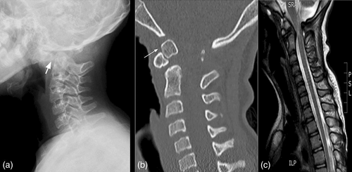
The autosomal dominant spondylometaphyseal dysplasia Sutcliff type or corner fracture type FN1-related is characterized by a combination of metaphyseal irregularities simulating fractures (“corner fractures”), developmental coxa vara, and vertebral changes. It is linked to heterozygous mutations in FN1 and COL2A1. Vertebral changes as delayed vertebral ossification, ovoid vertebral bodies, anterior vertebral wedging, and platyspondyly have been observed in this condition, while odontoid abnormalities have not been reported. We report an odontoid anomaly in a girl with SMD-CF FN1-related showing the heterozygous variant c.505T>A; p.(Cys169Ser), presenting at 11.9 years of age with acute quadriparesis. Images showed spinal cord compression and injury associated with os odontoideum and C1–C2 instability. She required decompression and instrumented occipitocervical stabilization, suffering from residual paraparesis. This paper describes the first case of SMD-CF FN1-related accompanied by odontoid anomalies.
1 INTRODUCTION
The autosomal dominant spondylometaphyseal dysplasia Sutcliff type or corner fracture type FN1-related (SMD-CF, MIM #184255) (Unger et al., 2023), was described as a separate entity by Langer et al. (1990). It is characterized by a combination of metaphyseal irregularities simulating fractures (“corner fractures”), developmental coxa vara, and vertebral changes (Langer et al., 1990). These “corner fractures”, described mainly in knees and wrists, are triangular bone fragments at the margin of the metaphysis that represent irregular ossification at the growth plates and secondary ossification centers, disappearing after growth has stopped (Currarino et al., 2000).
Classically it is described that the radiological findings in the metaphysis are visible at around 2 years of age, after children initiates walking and clinical findings as waddling gait, genu varum, and growth retardation appear (Langer et al., 1990).
It is linked to heterozygous mutations in FN1 (Cadoff et al., 2018; Costantini et al., 2019; Lee et al., 2017), although similar radiological findings were reported in individuals carrying heterozygous variants in COL2A1 (Machol et al., 2017; Walter et al., 2007).
Vertebral changes as delayed vertebral ossification, ovoid vertebral bodies, anterior vertebral wedging, and platyspondyly have been observed in SMD-CF as well as severe scoliosis and cervical kyphosis (Cadoff et al., 2018; Costantini et al., 2019; Currarino et al., 2000; Franceschini et al., 2005; Langer et al., 1990; Lee et al., 2017; Nair et al., 2016; Sutton et al., 2005). While odontoid abnormalities have not been reported in SMD-CF FN1-related, the absence of the odontoid process and vertebral compression due to subluxation of C1 in C2 was described in 1974 in an individual with SMD-CF (Felman et al., 1974).
We report an odontoid anomaly in a girl with SMD-CF FN1-related.
2 CASE DESCRIPTION
A girl presented at 11.9 years of age with acute quadriparesis after falling from her own height. Images showed spinal cord compression and injury associated with os odontoideum and C1–C2 instability (Figure 1a–c). She required decompression and instrumented occipitocervical stabilization, suffering from residual paraparesis.

She is the second daughter of a non-consanguineous healthy couple in a series of four. She was born premature due to uterine isthmus insufficiency, at 33 weeks of gestational age, with adequate size (weight 1320 g, −1.54 score standard deviation—SSD, length 42.0 cm, −0.76 SSD) (Villar et al., 2014).
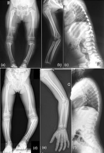
Her first visit was at 2.7 years old due to waddling gait and progressive genus varus noticed since the acquisition of gait, her length was −3.4 SSD. Radiographs showed metaphyseal defects at wrists, hips, knees, and ankles, with corner fracture images at medial sides of proximal tibias. In spine ovoid vertebral bodies (Figure 2). The progression of the lower limbs deformity required a tibial alignment osteotomy at 9.8 years old, it evolved with delayed tibial consolidation.

After obtaining informed consent, a Skeletal Dysplasia customized panel (SkeletalSeq V8) was performed, showing an heterozygous variant in exon 4 in FN1 (NM_212482.2): c.505T>A; p.(Cys169Ser). This missense type variant affects a highly conserved amino acid of the N-terminal assembly domain of FN1 and has been classified as deleterious by bioinformatic pathogenicity prediction tools. It was not present in population databases (gnomAD) nor in mutation databases (ClinVar, LOVD, and HGMD) at the time of the study. Another variant affecting the same amino acid, c.506G>A; p.(Cys169Tyr), was described in a patient with SMD-CF (Costantini et al., 2019). For all these reasons, it was classified as probably pathogenic following the ACMG variant classification recommendations (Richards et al., 2015).
3 DISCUSSION
The corner fracture type of autosomal dominant spondylometaphyseal dysplasia (SMD-CF) (MIM #184255) is an infrequent skeletal dysplasia, less than 20 cases of FN1 associated SMD-CF have been described in the literature (Cadoff et al., 2018; Costantini et al., 2019; Lee et al., 2017).
Clinical findings, such as waddling gait, genu varum, and growth retardation, and radiological findings in the metaphysis are visible at around 2 years of age (Langer et al., 1990). Our patient's first visit was at 2.7 years old, when waddling gait, progressive genus, and growth retardation were evidenced.
Vertebral changes, scoliosis, and kyphosis have been reported in SMD-CF (Cadoff et al., 2018; Costantini et al., 2019; Currarino et al., 2000; Franceschini et al., 2005; Langer et al., 1990; Lee et al., 2017; Nair et al., 2016; Sutton et al., 2005). Costantini et al. (2019) has described severe cervical kyphosis in a 59 years old woman with FN1 associated SMD-CF carrying a variant affecting the same amino acid as in this case c.506G>A; p.(Cys169Tyr), but odontoid abnormalities have not been reported in FN1 associated SMD-CF.
The absence of the odontoid process and vertebral compression due to subluxation of C1 in C2 was described by Felman in 1974 in an individual with SMD-CF, although this case did not have molecular confirmation, and odontoid hypoplasia was described in COL2 associated SMD-CF (Felman et al., 1974; Walter et al., 2007). This paper describes the first case of SMD-CF FN1-related accompanied by odontoid anomalies.
AUTHOR CONTRIBUTION
Rosario Ramos-Mejía: conception, data collection and analysis. Writing—original draft. Karen E. Heath: genetic analysis and test interpretation. Writing—review and editing. Silvia Modamio-Høybjør: genetic analysis and test interpretation. Writing—review and editing. Victoria Huckstadt: genetic analysis and test interpretation. Writing—review and editing. Julián Calcagni: selection and description of the images. Writing—review and editing. Rodrigo Remondino: selection and description of the images. Writing—review and editing. Virginia Fano: conception. Writing—review and editing. All the authors have been involved in drafting the manuscript or revising it critically for important intellectual content; and have given final approval of the version to be published.
Open Research
DATA AVAILABILITY STATEMENT
Data sharing is not applicable to this article as no new data were created or analyzed in this study.


