A patient with Cantú syndrome associated with fatal bronchopulmonary dysplasia and pulmonary hypertension
TO THE EDITOR
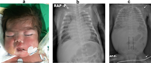
We describe a patient with Cantú syndrome confirmed by mutation analysis with associated cardiopulmonary complications. A female infant was born by vaginal delivery at 35 and 6/7 weeks of gestation, after a pregnancy complicated by polyhydramnios beginning at 32 weeks. She was the first child of nonconsanguineous Korean parents. Her birth weight was 3,240 g, length was 50 cm, and head circumference was 35 cm, all of which were >90th centile. Hypertrichosis was evident at birth. She had a coarse face with fullness of upper eyelids, a wide nasal bridge, and thick vermillion of the upper and lower lips (Fig. 1a). The initial chest radiograph showed relatively small lung volumes with mild cardiomegaly. On day 3, an echocardiogram showed a large patent ductus arteriosus (PDA) measuring 7 mm. It did not respond to medical treatment and was surgically ligated on the patient's seventh day of life. Repeat echocardiogram noted persistent pulmonary hypertension. Routine laboratory investigations were normal, including thyroid function tests. Her plasma and urine amino acid profile, lysosomal enzyme analysis and peripheral blood karyotype were normal. Ultrasonography of the head and abdomen and ophthalmological examination results were normal. A follow-up chest radiograph showed persistent cardiomegaly and worsening bronchopulmonary dysplasia (BPD) (Fig. 1b). However, despite gentle ventilatory support with steroid therapy, her BPD progressed, and a tracheostomy was performed at 82 days of age. Supplemental nitrous oxide gas was administered. A follow-up echocardiogram showed progressive pulmonary hypertension with tricuspid and mitral regurgitation and interventricular septal wall thickening (10 mm), but relatively good ventricular function (left ventricular ejection fraction, 75%). At 4 months of age, cranial magnetic resonance imaging showed atrophic changes of the brain. The baby's general condition continued to worsen and she developed recurrent and refractory pneumothoraces and sepsis. She eventually died of cor pulmonale, sepsis, and pneumothorax at 248 days of age. Genetic testing for Cantú syndrome was recommended by a clinical laboratory geneticist in the Netherlands who was consulted, and a heterozygous c.4385C>G (p.Ala1462Gly) mutation in ABCC9 was found postmortem. The alanine at position 1,462 is conserved from human to zebrafish and its substitution into glycine is predicted to have a deleterious effect on SUR2 protein function. After determination of the mutation, retrospective review of the infant's phenotype, cardiac manifestations, and radiologic findings were all consistent with Cantú syndrome. We then conducted mutation analysis and biologic parentage analysis. Neither parent had the pathogenic mutation in the ABCC9. Biologic parentage analysis confirmed that they are truly biologic parents of the patient. We conclude that, the mutation of the patient likely has been arisen de novo in the proband and was the cause of the Cantú syndrome. The authors declare that these studies are not considered to be research at their institution.

Cantú syndrome is a rare disorder comprising congenital hypertrichosis, distinctive facial features, osteochondrodysplasia, and cardiomegaly and was first described by Cantú et al. [1982]. Only approximately 40 patients with Cantú syndrome have been reported [Nevin et al., 1996; García-Cruz et al., 1997; Rosser et al., 1998; Robertson et al., 1999; Concolino et al., 2000; Lazalde et al., 2000; Engels et al., 2002; Tan et al., 2005; Grange et al., 2006; García-Cruz et al., 2011; Kurban et al., 2011; Scurr et al., 2011; Harakalova et al., 2012; van Bon et al., 2012].
Hypertrichosis and dysmorphic facies are observed in most patients with Cantú syndrome and are usually evident at birth. Affected infants have thick scalp hair extending onto the forehead and heavy, generalized body hair. A coarse facial appearance is also typical, with a wide or depressed nasal bridge, epicanthal folds, and a wide mouth with thick vermillion of the lips. In addition, macrosomia and macrocephaly are also frequently observed.
Grange et al. [2006] reviewed the cardiac manifestations of Cantú syndrome and suggested that cardiomegaly seems to be caused by increased cardiac muscle mass, not by a true cardiomyopathy. Most reported patients have shown normal ventricular function. A PDA is the only significant reported structural heart defect, and surgical ligation is needed in some cases. Pulmonary hypertension has also been observed in patients with Cantú syndrome, as in the patient described here. However, the cause of pulmonary hypertension in Cantú syndrome is unknown, and its long-term outcome cannot be predicted. Scurr et al. [2011] reported a patient with pulmonary hypertension who appeared to respond to steroid therapy and gradually improved; they suggested the possibility of inflammation as an underlying pathology of Cantú syndrome. However, no reported patient has been as severely affected as the patient reported here, and this patient's pulmonary hypertension was not responsive to repeated steroid therapy. We could not determine the underlying pathphysiology of the patient's pulmonary hypertension because we did not perform cardiac catheterization. Now that the genetic cause of Cantú syndrome is known, some of the manifestations may be explained as a consequence of ABCC9 dysfunction, although this does not explain all of the manifestations. Furthermore, the clinical course of the present patient was significantly different from that of the previously reported patients. It is not possible for us to determine if the cardiopulmonary complications are a direct consequence of the Cantú syndrome and ABCC9 dysfunction or if they are coincidental. Additional reports are necessary to affirm or refute this association.
The known radiological features of osteochondroplasia in Cantú syndrome are a narrow thorax, wide ribs, coxa valga, osteopenia, enlarged medullary canals with an “Erlenmeyer-flask” appearance, and metaphyseal widening of long bones [Rosser et al., 1998; Concolino et al., 2000]. Other described skeletal abnormalities include flattened or ovoid vertebral bodies, thickened calvaria, growth arrest in the metaphyses, and small terminal phalange of the thumbs and halluces [Engels et al., 2002; Czeschik et al., 2013]. The patient reported here had no features of osteochondrodysplasia of Cantú syndrome at birth; however, at 2–3 months of age, some characteristic findings were observed on radiologic examination, including wide ribs, delayed ossification of the humeral and femoral epiphyses, and narrow interpedicular distance of the caudal lumbar spine (Fig. 1c).
Heterozygous missense mutations of ABCC9 cause Cantú syndrome [Harakalova et al., 2012]. The SUR2 protein encoded by ABCC9 is part of an ATP-sensitive potassium channel complex (ATP-sensitive meaning that KATP channels open and close in response to the intracellular ADP/ATP ratio) that functions primarily in the heart, skeletal muscle, and smooth muscle. The ABCC9 mutations may disturb the structure, thereby leading to dysfunction of the KATP channels. By electrophysiological measurements, ABCC9 mutations were found to reduce the ATP-mediated potassium channel inhibition, resulting in channel opening and eventually leading to the various clinical features of Cantú syndrome [Harakalova et al., 2012]. Cantú syndrome is a channelopathy and further understanding of the underlying pathologic mechanism may lead to effective therapeutic options [Bregje et al., 2012].


