Cervical vertebrae, cranial base, and mandibular retrognathia in human triploid fetuses†
How to cite this article: Sonnesen L, Nolting D, Engel U, Kjær I. 2009. Cervical vertebrae, cranial base, and mandibular retrognathia in human triploid fetuses. Am J Med Genet Part A 149A:177–187.
Abstract
On profile radiographs of adults, an association between fusions of cervical vertebrae, deviations in the cranial base and mandibular retrognathia has been documented radiographically. An elaboration of this association on a histological level is needed. In human triploid fetuses severe mandibular retrognathia and deviations in the cranial base have previously been described radiographically (without cephalometry) and cervical column fusions radiographically as well as histologically. Therefore, triploid fetuses were chosen to elucidate the cranial base cephalomterically and histologically. In the present study, eight triploid fetuses were analyzed radiographically and histologically focusing especially on the cranial base, which borders to the spine and to which the jaws are attached. A histological analysis of the cranial base has not previously been performed in triploid cases. An enlarged cranial base angle and a retrognathic position of the mandible were documented cephalometrically on radiographs of all cases. Histologically, malformations were observed in the cranial base as well as in the spine. These are new findings indicating the association between the occipital bone and the uppermost vertebra in the body axis. As the notochord connects the cervical column and the cranial base in early prenatal life, molecular signaling from the notochord may in future studies support the notochord as the developmental link between abnormal development in the spine and the cranial base. © 2009 Wiley-Liss, Inc.
INTRODUCTION
In human triploid fetuses severe retrognathia of the mandible has been described [Schrocksnadel et al., 1982; Bareggi et al., 2001], and malformation of the cervical vertebrae has been documented radiographically and histologically [Nolting et al., 1998, 2002]. Thus, triploid fetuses may serve as a model for elucidating associated anomalies in the cervical column, cranial base and craniofacial profile. The bones surrounding the most cranial part of the notochord, which is the cranial base between the sella turcica and the foramen magnum [Hamilton and Mossman, 1972; Hanken and Hall, 1993; Kjær et al., 1999], have not previously been analyzed in triploid fetuses.
The aims of the present study were: (1) To describe the cervical column and the cranial base between the sella turcica and the foramen magnum in human triploid fetuses on radiographs and to verify the findings histologically; (2) to compare the findings in the cervical column with the findings in the cranial base and (3) to perform a cephalometric analysis in human triploid fetuses and compare the results with normal reference values.
MATERIALS AND METHODS
We studied eight human triploid fetuses from spontaneous and induced abortions after karyotyping at the Hvidovre University Hospital, Copenhagen, Denmark. The fetuses were examined radiographically and histologically in accordance with legal autopsy procedures including parental consent for autopsy of the central nervous system (CNS).
Two fetuses had the karyotype 69,XXY and 6 were 69,XXX. Gestational ages were 15–23 weeks, CRL 97–190 mm (Table I). Gross malformation of the CNS was not registered in any of the cases.
| GA (weeks) | CRL (mm) | Karyotype | Radiography | Histology | Illustration figure | |||
|---|---|---|---|---|---|---|---|---|
| Column | Cranial base | Jaws | Column | Cranial base | ||||
| 15 | 97 | 69,XXX | + | + | + | − | + | 5, 9 |
| 16 | 125 | 69,XXX | + | + | + | + | + | 1, 2, 5 |
| 18 | 160 | 69,XXX | + | + | + | + | + | 3 |
| 19 | 105 | 69,XXX | + | + | + | + | + | 8 |
| 20 | 110 | 69,XXX | + | + | + | − | + | |
| 20 | 152 | 69,XXY | + | + | + | − | + | 1 |
| 21 | 100 | 69,XXY | + | + | + | + | + | 6, 7 |
| 23 | 190 | 69,XXX | + | + | + | + | + | 4, 10 |
- +, available for the study; −, not available for the study.
Radiographic Method
In each of the eight fetuses the cervical column and the craniofacial region were radiographed in frontal, lateral and axial projections. The specimens were fixed and orientated on a Plexiglas plate placed directly on the film before radiography. For radiography, a Grenz ray radiographic apparatus (Hewlett Packard Faxitron, Model 43855A, McMinniville, OR) was used. The tube voltage varied between 30 and 60 kV and the exposure time from 10 to 60 sec at 2.8–3.0 mA, depending on the size of the specimen. The film used was Kodak X-omat MA (Kodak-Industrie, Marne-La-Valle, France), routinely processed. The results were compared with those of normal vertebral and cranial development described previously [Kjær, 1990; Kjær et al., 1993; Kyrkanides et al., 1993]. Fusion and proportion of the cervical column was described. Fusion in the spine was described as fusion between vertebrae in the region of the articulation facet, neural arch or transverse processes [Sonnesen et al., 2008]. Cervical disproportion was described as a lack of proper relationship between parts of vertebrae.
Cephalometric Analysis
-
ba, basion—the most caudal point of the lower border of the occipital corpus.
-
s, sella—the constructed midpoint of the sella turcica, which is the midpoint of a line connecting the top of the dorsum sellae to the midpoint of the upper contour of the ossified part of the post-sphenoid bone [van den Eynde et al., 1992].
-
n, nasion—the point of intersection between a line tangential to the nasal bone and a line tangential to the lower 5 mm of the frontal bone.
-
sp, spinal point—the apex of the anterior nasal spine.
-
pg, pogonion—the most prominent point of the mandible. The measurements were compared with normal values previously described by van den Eynde et al. 1992.

The prenatal craniofacial profile. Upper left: Cephalometric reference points in a normal prenatal craniofacial profile according to van den Eynde et al. 1992. Red line indicates the course of the cranial end of the notochord connecting the vertebral bodies and cranial base. Green star indicates the basilar part of the occipital bone. Lower left: Illustration of the normal prenatal contour of the basilar part of the occipital bone, approximate age 19 weeks. Yellow star indicates the frontal view, and green star indicates the lateral view. Upper right: Profile radiograph of a human triploid fetus, gestational age 16 weeks. X1: Green star indicates basilar part of occipital bone. Lower right: Profile radiograph of a human triploid fetus, gestational age 20 weeks. X1: Green star indicates basilar part of occipital bone.
Histological Method
After radiography, midaxial cranial base tissue blocks from the sella turcica (including the pituitary gland) to the foramen magnum and tissue blocks including the cervical vertebrae were examined histochemically. These tissue blocks were fixed in 10% neutral buffered formalin, demineralized 2–20 days in equal parts of 2% citric acid and 20% sodium citrate (pH 6), embedded in paraffin, serially cut into 4–6 µm sections, and stained with toluidine blue pH 7 [Nolting et al., 1998].
RESULTS
Cervical Column
In all cases, malformation of the cervical spine was found on radiographs (Table II). Four fetuses had fusion of 2–5 cervical vertebrae (Table II and Fig. 2), seven fetuses had disproportion of the cervical vertebral bodies (Table II and Fig. 3). Three fetuses had fusion as well as disproportion of the vertebrae (Table II and Fig. 4).
| GA weeks | Malformation (X-ray) | Basilar part of occipital bone | Malformation (X-ray) | Postsphenoid bone | Cervical column malformation (X-ray) | |||
|---|---|---|---|---|---|---|---|---|
| Trabeculae | Cortical bone | Shape | Subpharyngeal | Sella turcica morphology | ||||
| 15 | + | Few | Malformed | Extremely slender | Bilateral centers | Concavity | Recess anterior | Disproportion minor |
| 16 | + | Few | Malformed | Slender, cleft | Bilateral centers | Minor concavity | Wide cranial | Fusion |
| 18 | + | — | — | — | Partial cleft | Normal | Recess anterior | Disproportion, fusion |
| 19 | + | Few | Malformed | Slender | Bilateral centers | Concavity | Recess anterior | Disproportion |
| 20 | + | Few | Normal | Slender | Partial cleft | — | Recess anterior | Disproportion, minor |
| 20 | + | Few | Malformed | Slender | Partial cleft | Concavity | Recess anterior | Disproportion |
| 21 | + | Few | Malformed | Extremely slender | Bilateral centers | Concavity | Normal | Disproportion, fusion |
| 23 | + | — | — | — | Partial cleft | Normal | Wide cranial | Disproportion, fusion |
- Radiography and histology.

The cervical column in a human triploid fetus, gestational age 16 weeks. a: Radiograph in frontal projection. X2: Marked area: vertebral body fusion of C2-C6. b: Radiographs in lateral projection. X2: (c) Histological frontal section of vertebral body fusion of C2–C5. Ossification occurs midaxially in the cartilaginous vertebral bodies and between the cartilaginous anlagen. Toluidine blue. X5: The marked area is shown in (d). d: Magnification of the ossified area demonstrated in (c). In the cartilage changes in the metachromasia are observed in the most axial part indicated by arrows. This change in metachromasia is located in the original notochordal region. X24.

The cervical column in a human triploid fetus, gestational age 18 weeks. a: Radiograph in frontal projection. Marked area: vertebral body disproportion of C2–C6. Red star indicates partial cleft of the postsphenoid bone. Yellow star indicates basilar part of the occipital bone, malformed in the lower aspect. X1.5. b: Radiograph in lateral projection. c: Histological frontal section of vertebrae marked in (a). Arrow indicates nucleus pulposus disci invertebralis. Toluidine blue. X4.

The cervical column in a human triploidy fetus, gestational age 23 weeks. a: Radiograph in frontal projection. Marked area: vertebral body disproportion of C4-T2. Vertebrae fusion between C7 and T1. Yellow stars indicate complete cleft of the basilar part of the occipital bone. X1.5. b: Radiograph in lateral projection. X1.5. c: Histological frontal section of area marked in (a). Arrow indicates distinct metachromatic “borders” between cartilage in the original notochordal region and the surrounding cartilage. Toluidine blue. X4.
In five cases, it was possible to analyze the vertebral column histologically, showing that in cases with fusion, midaxial ossification occurred within the cartilaginous vertebral bodies and between these cartilage anlagen (Fig. 2c). In cases with disproportions, different sizes of the vertebral bodies were observed (Fig. 3c). Thus, the radiographic findings of bone malformations were confirmed histologically (Figs. 2-4).
Also, the cartilage appeared different in the vertebral column. The outer contour of the individual cartilaginous corpora was morphologically normal in all cases compared with normal standards, while marked changes in metachromasia were seen in the most axial part (Figs. 2d and 4c). Distinct “borders” were observed between these central areas located in the original notochordal regions and the surrounding cartilage (Figs. 2d and 4c).
The nucleus pulposus disci invertebralis was visible in areas without body fusions but not in areas with fusions (Figs. 2c and 3c).
Cranial Base
The bony elements identified in the cranial base blocks were the basilar part of the occipital bone and the postsphenoid bone including the sella turcica. Malformations in the cranial base occurred in all cases (Table II).
Radiography of the Cranial Base
The basilar part of the occipital bone was affected in all cases (Table II). In six cases, concavities in the anterior part of the bone, either unilateral or bilateral, were found on frontal projection (Figs. 5 and 6), and in one case malformation of the posterior part was observed (Fig. 3a). In one case a complete cleft of the basilar part of the occipital bone was observed in the frontal plane (Fig. 4a).

Radiographs in axial projection of the cranial base structures in two human triploidy fetuses, gestational ages 15 (left, X2) and 16 weeks (right, X1.3). Red stars indicate bilateral ossification centers of the postsphenoid bone. Yellow stars indicate the basilar part of the occipital bone. Note concavity in the anterior part.

Radiograph in frontal projection of the cervical column and posterior part of the cranial base in a human triploid fetus gestational age 21 weeks. Red stars indicate bilateral ossification centers of the postsphenoid bone. Yellow star indicates the basilar part of the occipital bone. Note concavity in the anterior part. Vertebral body fusion of C6-T2 and vertebral body disproportion of C1–C5 in the cervical column. X2.
The postsphenoid bone was also affected in all cases (Table II). Bilateral ossification centers were observed in four cases (Figs. 5 and 6), and a partial cleft was observed in four cases (Fig. 3a).
Histologic Findings of the Cranial Base
In two cases, it was not possible to analyze the basilar part of the occipital bone due to disruption of the tissue during autopsy. In the remaining six cases, the basilar part of the occipital bone appeared slender, in two of these cases extremely slender (Figs. 7-9), and in one of the six cases the basilar part of the occipital bone was completely cleft. Deficiency or absence of the cortical bone was observed (Figs. 7-9), and the inner bony contour was characterized by few trabeculae compared with normal conditions (Figs. 7-9). The basilar part of the occipital bone was cranially positioned, and the cartilage located anterior to this bone was thin and often with a deep concavity in the cranial contour (Figs. 8 and 9).

Sagittal histological sections of the basilar part of the occipital bone and the postsphenoid bone including the sella turcica in a human triploid fetus, gestational age 21 weeks. a–c: Arrows in a and b indicate deficiency or absence of the cortical bone in the basilar part of the occipital bone. The inner bony contour is characterized by few trabeculae. In c open arrow indicates the most cranial remains of the notochord located in the cartilage posteriorly to the sella turcica. Toluidine blue. X5.5. d: Magnification of marked area in (c). Note the interruption of bony contour due to the anterior cavities in the basilar part of the occipital bone demonstrated radiographically in Figure 6. Toluidine blue. X14.

Sagittal histological section of the basilar part of the occipital bone and the postsphenoid bone including the sella turcica in a human triploid fetus, gestational age 19 weeks. Black star indicates recess in the anterior wall of the sella turcica, postsphenoid bone. Open arrow indicates concavity of the postsphenoid cartilage and bone subpharyngeally. Arrow indicates a deep contour in the cartilage located anterior to the basilar part of the occipital bone. Toluidine blue. X10. Inserted figure in upper right corner shows few trabeculae in the basilar part of the occipital bone (marked below). Toluidine blue. X18.

Sagittal histological section of the basilar part of the occipital bone and the postsphenoid bone including the sella turcica in a human triploid fetus, gestational age 15 weeks. Black star indicates recess anterior at the base of the sella turcica, postsphenoid bone. Open arrow indicates concavity of the postsphenoid cartilage and bone subpharyngeally. Open arrow indicates a deep contour in the cartilage located anterior to the basilar part of the occipital bone. Few trabeculae are seen in the basilar part of the occipital bone. Toluidine blue. X13. Inserted figure in upper right corner shows the sella turcica from a neighboring section from the same specimen. Note the deep recess anterior at the base of the sella turcica. Arrow indicates remnants of the most cranial part of the notochord located in the dorsum sella. Toluidine blue. X13.
In the postsphenoid bone, abnormal sella turcica morphology was observed in all cases except one. In seven cases, the deviation in the sella turcica morphology varied (Figs. 8 and 9). In four cases, malformation of sella turcica was seen as a recess anterior to the inferior of the sella (Fig. 9) and in one case as a recess in the anterior wall (Fig. 8). In two cases, the sella turcica was wider cranially than seen under normal conditions (Fig. 10). In five cases, a concavity of the postsphenoid cartilage and bone was observed subpharyngeally (Figs. 8 and 9).

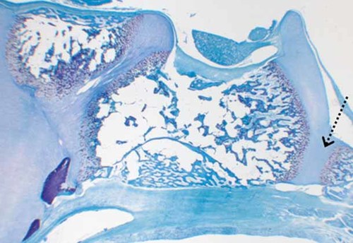
Sagittal histological section of the postsphenoid bone and the border between the basilar part of the postsphenoid bone and the occipital bone in a human triploid fetus, gestational age 23 weeks. Dotted arrow indicates the spheno-occipital synchondrosis with marked differences in the metachromasia of cartilage peripherally and centrally in the synchondrosis. Toluidine blue. X5.
The most cranial part of the notochord was identified midaxially in the dorsum sellae in seven cases (Figs. 7c and 9).
The cartilage between the basilar part of the occipital bone and the postsphenoid bone (the developing sphenobasilar synchondrosis) deviated in two cases. In these two cases differences in cartilage metachromasia were observed (Fig. 10).
Comparison of the Findings in the Cervical Column and Cranial Base
In all cases, malformation of the cervical column and of the cranial base was observed (Fig. 6). The malformation of the cervical spine and of the cranial base occurred in bony and cartilaginous tissue. It was not possible to classify the cranial base malformations according to a specific pattern of column malformations (Table II).
Cranial Base Angle and Prognathia of the Jaws
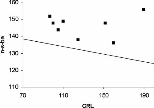
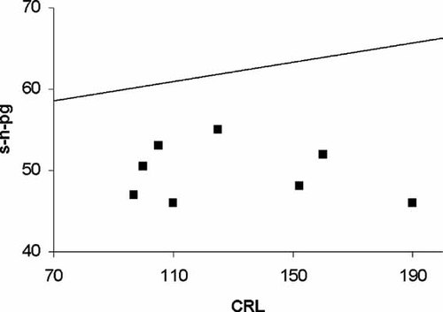
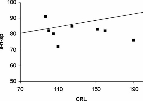
In all fetuses, the cranial base angle (n-s-ba) was larger compared to controls (Figs. 1 and 11). Also, in all fetuses the mandible was more retrognate (s-n-pg) compared with controls (Figs. 1 and 12). The prognathia of the maxilla (s-n-sp) was approximately the same compared with the control material (Figs. 1 and 13).

Cephalometric values of cranial base angle (n-s-ba) (black squares) in human triploid fetuses compared with a line representing normal mean values according to van den Eynde et al. 1992. Crown rump length (CRL). All triploid specimens had a larger cranial base angle compared with normal mean values.

Cephalometric values of the mandibular prognathia (s-n-pg) (black squares) in human triploid fetuses compared with a line representing normal mean values according to van den Eynde et al. 1992. Crown rump length (CRL). All triploid specimens had mandibular retrognathia compared with normal mean values.

Cephalometric values of the maxillary prognathia (s-n-sp) (black squares) in human triploid fetuses compared with a line representing normal mean values according to van den Eynde et al. 1992. Crown rump length (CRL). The prognathia of the maxilla was about the same compared with the control material.
DISCUSSION
We have studied eight human triploid fetuses, triploidy being defined as the presence of three haploid sets of chromosomes to a total of 69 chromosomes, the sex chromosomes most often being either XXX or XXY [Carr, 1971; Gilbert and Opitz, 1982]. Triploidy is known to be one of the most frequent chromosome abnormalities in first-trimester abortions, accounting for 18% of spontaneous miscarriages and occurring in 1% of all recognized pregnancies [Niemann-Seyde et al., 1993]. The condition is lethal within the first hours after birth and the longest survival for a full triploid infant was reported as 10.5 months [Niemann-Seyde et al., 1993].
One of the aims of the present study was to analyze the cranial base, which has never been analyzed in triploid fetuses. It is interesting to study the cranial base because caudally it borders on the vertebral column and cranially it is connected to the craniofacial skeleton. Since neural tube malformations such as anencephaly and myelomeningocele influence the cranial base angulation [Kjær et al., 1994a,b, 1998], we excluded triploid fetuses with such malformations. Failure in closure of the neural tube during early embryonic life results in a bent cranial base and a small cranial base angle.
Cervical Column
All examined tissue blocks of the vertebrae showed malformations such as fusion or disproportion in size. The histological findings of the tissue blocks confirm the radiological findings. This is in agreement with a previous study [Nolting et al., 2002]. The term fusion is used for the morphological appearance of united vertebral bodies. This morphological appearance may be explained by an early embryonic defect in the segmentation of the column, that is, non-segmentation [Kjær et al., 1997b]. In the present study, it was found that nucleus pulposus disci intervertebralis was lacking in areas with vertebral fusions. The nucleus pulposus is a semifluid mass of fine elastic fibers that forms the central portion of an intervertebral disc. It has been regarded as the persistent remains of the embryonic notochord [Hamilton and Mossman, 1972; Lohse et al., 1985; Lammerding-Koppel et al., 1995; Smits and Lefebvre, 2003; DiPaola et al., 2005; Stemple, 2005]. Thus, fusion of vertebral bodies may represent a notochordal malfunction. Fusions of vertebral bodies in early life could also be one explanation why crown rump length is severely reduced for age in human triploid fetuses [Gilbert and Opitz, 1982; Bareggi et al., 2001].
It is not possible to determine whether the fusion seen in prenatal life is of the same type of fusion seen in postnatal life [Sonnesen et al., 2007]. The fusions observed prenatally may impair growth in the vertebral column at an early stage. It could be argued that postnatal fusion of vertebral bodies appears after gradual degeneration of the nucleus pulposus and the surrounding inter-vertebral tissues and is gradually replaced by cartilage and bone.
Cranial Base
Histological analyses of the cranial base have been performed previously in normal fetuses [Kyrkanides et al., 1993]. We analyzed cephalometrically and histologically the cranial base in human triploid fetuses, which has not previously been reported. In the present study, malformation of the cervical column and of the cranial base was found in all cases. The skull including the cranial base has a complex phylogenetic history, which is associated with its progressive modification in vertebrate phylogeny and with the adaptive specialization of many of its components [Hamilton and Mossman, 1972; Hanken and Hall, 1993]. According to Hamilton et al., the development of the cranial base occurs in the parachordial region in close relation with the cephalic part of the notochord, that is, from the region of the caudal end of the occipital corpus to the hypophysis and a prechordal or trabecular region in front of the notochord [Hamilton and Mossman, 1972]. Previously, prenatal studies have shown a notochordal connection between the cervical vertebrae and the posterior part of the cranial base [Hamilton and Mossman, 1972; Müller and O'Rahilly, 1980; Hanken and Hall, 1993; Kjær, 1995, 1998; Kjær and Hansen, 1995; Kjær et al., 1994a, 1999; Sadler, 2005]. Thus, the notochord has a direct connection to the basilar part of the occipital bone, but only a hypothetical connection to the neural crest. Whether the occipital bone can be considered the uppermost vertebra in the body axis, has been discussed extensively in the literature since 1790 [de Beer, 1971; Hanken and Hall, 1993]. In the present study, we observed that the occipital bone was malformed in triploidy with severe spine malformations. This leads to the hypothesis that the occipital bone is formed and patterned by the same developmental mechanism as the vertebral column, thus supporting the theory that the occipital bone is considered the uppermost vertebral bone. The developmental connection between the spine and the occipital bone has recently been illustrated by in situ marking of the body axis in mice embryos using Collagen II, Pax9, Pax1, and Noggin antibodies [Sonnesen et al., 2008]. This study showed a distinct Pax1 expression in the spine as well as in the basilar part of the occipital bone. No expression was seen in the craniofacial region. Meanwhile, Pax9 was expressed in the spine and in the craniofacial skeleton, and only vaguely in the cranial base.
A deviation in the development of the notochord or the signaling from the notochord may influence the surrounding bone tissue in the spine and in the posterior part of the cranial base as seen in the present study. However, it was not possible to classify the cranial base malformations according to a specific pattern of column malformations, in part due to small numbers in the present study. The axial segmental differences in expression of HOX genes may explain why some spine segments are malformed, while others are not [Kjær et al., 1996, 1997a; Keeling et al., 1997; Nolting et al., 2000].
Cranial Base Angle and Prognathia of the Jaws
The cranial base angle and the prognathia of the mandible and maxilla in triploid human fetuses have not previously been measured cephalometrically or compared to normal values. In the present study, a large cranial base angle was found in all human triploid fetuses compared with previously described normal values [van den Eynde et al., 1992]. This is in agreement with the present study of the position of the basilar part of the occipital bone. As the basilar part of the occipital bone was cranially positioned, the basion point has a more cranial location resulting in an enlarged cranial base angle.
We found severe mandibular retrognathia in all human triploid fetuses compared to normal values [van den Eynde et al., 1992]. Postnatally, retrognathia of the mandible was reported in the Klippel-Feil anomaly [Smith and Griffin, 1992] and in Ullrich Turner syndrome in girls [Jensen, 1985] and in human fetuses [Andersen et al., 2000]. In the postnatal conditions, fusions in the cervical column were reported.
Retrognathia of the fetal mandible described in the present study could be a manifestation associated with the enlarged cranial base angle. Previously, it was noted that a large cranial base angle in children and adults results in retrognathia of the mandible [Björk, 1947, 1975]. The severe mandibular retrognathia in the present study may be a result of abnormal signaling from the notochord to the neural crest cells determining the craniofacial morphology before the notochord is surrounded by bone tissue and disappears [Müller and O'Rahilly, 1980; Kjær, 1995, 1998; Sadler, 2005]. This is a hypothesis, which has still not been verified. As the mandible develops from Meckel's cartilage, deviations in the cartilaginous tissue as seen in the vertebral bodies in the present study could also explain the reduced size and position of the mandible [Kjær, 1997].
On profile radiographs, used for diagnosis in an orthodontic clinic, a correlation was demonstrated previously between the cervical column, cranial base and craniofacial profile [Sonnesen and Kjær, 2007a,b, 2008]. An explanation for this interrelationship has never been given. The present study demonstrates the value of performing histological analyses on fetal material in order to contribute to an explanation of the associations registered radiographically. With a view back to early notochordal location in humans and to the immunomarkings of body axis in mice, a genetically determined connection between the cervical column and the cranial base is suggested. The molecular connections between the spine, the cranial base and the craniofacial skeleton have previously been illustrated also by immunohistochemical analyses [Sonnesen et al., 2008]. The study showed that Pax9 is expressed in the spine and in the craniofacial skeleton thus suggesting a connection between the spine and the craniofacial region. Besides using Pax9 it would be appropriate to use markers such as SHH as potential signaling molecules and SOX5 and SOX6 as markers of notochordal differentiation.
Future molecular studies may further elucidate the importance of the notochordal function for the development of the cervical column, cranial base and craniofacial profile.
Acknowledgements
Birgit Fischer Hansen, MD and Jean W. Keeling, MD are acknowledged for excellent guidance in fetal pathology. Maria Kvetny, MA is acknowledged for linguistic support and manuscript preparation. The IMK Foundation is acknowledged for funding.


