Accurate identification of a precursor B-cell neoplasm
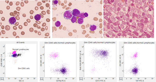
A 23-year-old man with a history of Crohn's disease and liver transplantation for sclerosing cholangitis developed pancytopenia during corticosteroid therapy. His blood count showed hemoglobin concentration 81 g/L, white cell count 7.1 × 10⁹/L, neutrophils 0.7 × 10⁹/L, and platelets 34 × 109/L. His blood film showed a population of medium sized lymphoid cells with a high nucleocytoplasmic ratio, some with prominent nucleoli and vacuolation (top left, all images ×100 objective). A large population of these cells were noted in the bone marrow aspirate (top center) and trephine biopsy sections (top right). Immunophenotyping of the CD45weak cells (purple events plot 1) showed these cells to express CD19, CD79a (plot 2), CD22, HLA-DR, and CD15 (plot 3). Importantly, CD34, CD117, myeloperoxidase, CD10, cytoplasmic CD3, CD79b, and terminal deoxynucleotidyl transferase (TdT) (plot 4), were not expressed, nor was cytoplasmic or surface membrane immunoglobulin (Ig).
The morphology in this case is indicative of a precursor neoplasm and B lineage was indicated by expression of CD19, CD79a, and CD22. Unusually CD34 and TdT were not expressed. The lack of surface and cytoplasmic Ig, weak CD45, absent CD79b and aberrant expression of CD15 are, however, all features of a precursor B-cell neoplasm. Pro-B acute lymphoblastic leukemia, despite arising from an early precursor, may fail to express TdT and sometimes CD34.1 It is often associated with a KMT2A translocation and this was shown in this case with t(4;11)(q21;q23.3) confirming the diagnosis. Furthermore, the glucocorticoid therapy used in the management of Crohn's disease in this patient may have induced downregulation of precursor antigen expression similar to that reported during induction chemotherapy.2, 3
CONFLICT OF INTEREST STATEMENT
The authors declare no conflicts of interest.


