Giant proerythroblasts in pure red cell aplasia due to parvovirus B19 infection in a patient with rheumatoid arthritis
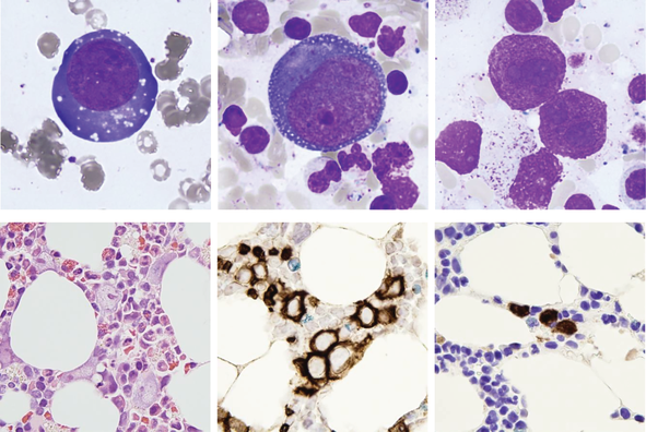 A 72-year-old woman with rheumatoid arthritis presented with fatigue and chest pain and was noted to have a hemoglobin concentration of 43 g/L with absolute reticulocytopenia (3 × 10⁹/L, normal range 50-100). The white cell and platelet counts were normal. The blood film showed no specific features. A bone marrow aspirate showed an absence of normal erythroid precursors with normal granulopoiesis and megakaryopoiesis. There were a number of extremely large cells up to 50 μm in diameter with deeply basophilic cytoplasm, occasionally with vacuolation (top left and center, ×100 objective). Some nuclei had inclusions and multiple bare nuclei from these giant cells were also apparent (top right, ×100). The bone marrow trephine biopsy confirmed the presence of giant proerythroblasts with an absence of more mature erythroid precursors (hematoxylin and eosin, ×50, bottom left; immunoperoxidase for glycophorin A, ×50 bottom center). Immunoperoxidase for parvovirus viral capsid antigens VP1 and VP2 showed positivity in the nuclei of the giant proerythroblasts (×50, bottom right). No parvovirus B19 IgM or IgG antibodies were detected but PCR for parvovirus DNA was positive.
Parvovirus B19 is a single stranded DNA virus that specifically infects red cell precursors. Pure red cell aplasia caused by this virus is associated with the presence of giant proerythroblasts, often with viral inclusions (“lantern cells”) in the bone marrow. This diagnosis should be considered in immunosuppressed individuals with apparent red cell aplasia as protracted infection and severe anemia can result from their inability to clear the virus. Serology may be negative and PCR analysis is often required for detection of the virus in these patients. The patient reported here was receiving methotrexate, sulphasalazine, hydroxychloroquine and golimumab (anti-TNFα) therapy for her rheumatoid arthritis. Treatment with intravenous immunoglobulin resulted in reticulocytosis within 2 weeks of administration.
1 CONFLICT OF INTEREST
The authors declare no potential conflict of interest.