Volume 94, Issue 11 pp. 1288-1289
CLINICAL PEARLS IN BLOOD DISEASES
Free Access

It's a black day—metastatic melanoma in the bone marrow

Barbara J. Bain

Corresponding Author

Barbara J. Bain

Department of Haematology, St. Mary's Hospital, London, UK

Centre for Haematology, St. Mary's Hospital Campus of Imperial College Faculty of Medicine, St. Mary's Hospital, London, UK

Correspondence

Barbara J. Bain, Department of Haematology, St Mary's Hospital, Praed Street, London, W2 1NY, UK.

Email: [email protected]

Search for more papers by this author
John Luckit

John Luckit

Department of Haematology, North Middlesex University Hospital, London, UK

Search for more papers by this author
First published: 04 August 2019
Citations: 1
When metastatic cells are present in the bone marrow it is rarely possible to identify the cellular origin of a tumor from morphology alone. An exception is neuroblastoma, when neurofibrils are detected.1 It can also be possible to suspect small cell carcinoma of the lung when the characteristic molding of nuclei is noted, and adenocarcinoma in general when secretory globules are present within tumor cells. One tumor that can be identified with certainty is malignant melanoma. These images are from a 33-year-old man who presented with right axillary lymphadenopathy. He had had a mole on his right shoulder from birth, which had changed characteristics in the preceding year.

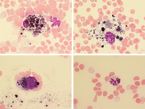
A bone marrow aspirate showed abundant macrophages containing melanin (Images, top). In addition there were melanoma cells (Image, bottom left), which were sometimes difficult to distinguish from macrophages. Neutrophils in the bone marrow contained melanin (Image, bottom right). The patient's trephine biopsy specimen was black on macroscopic examination. A buffy coat preparation from the peripheral blood of this patient has previously been reported; both circulating melanoma cells and neutrophils containing melanin were detected.2

A diagnosis of metastatic melanoma can be confirmed by immunohistochemistry but in this case the clinical and morphological features were sufficient for an unequivocal diagnosis.

CONFLICT OF INTEREST

Nothing to report.

    The full text of this article hosted at iucr.org is unavailable due to technical difficulties.