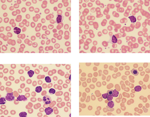
Diagnosis of follicular lymphoma from the peripheral blood
The bottom left image is from another middle aged man with lymphadenopathy, mild anemia (Hb 128 g/L), a normal platelet count, and a lymphocyte count of 89 × 109/L.
The blood film showed small and medium sized lymphocytes with dense chromatin; some were nucleolated and some had deep narrow nuclear clefts. Immunophenotyping showed clonal B cells expressing FMC7 and strong kappa light chain and not expressing CD5 or CD10; follicular lymphoma was confirmed on a lymph node biopsy.
The bottom right image is from a young woman with mild splenomegaly, a normal Hb and platelet count, and a lymphocyte count of 11 × 109/L. In addition to some nucleated red blood cells, there were small lymphocytes with similar features to those described in the first two patients. Clonal B cells expressed CD19, CD10, CD79b, FMC7, and strong kappa light chain with CD5 and CD23 being negative. The diagnosis of follicular lymphoma was confirmed on biopsy.
The blood film features most suggestive of follicular lymphoma are the presence of small lymphocytes with chromatin that is more uniformly condensed than that of chronic lymphocytic leukemia cells, with some cells showing deep, narrow nuclear clefts and with smear cells not being prominent. Immunophenotyping typically shows expression of pan-B markers with strong surface membrane immunoglobulin and no expression of CD23 or CD200; when there is some degree of pleomorphism, CD5 negativity helps to exclude mantle cell lymphoma and, when it is positive, CD10 expression supports the diagnosis of follicular lymphoma. This diagnosis can be confirmed by fluorescence in situ hybridization on peripheral blood cells, as well as by lymph node biopsy.
CONFLICT OF INTEREST
None.


