Volume 89, Issue 7 pp. 787-788
Diagnostic Imaging in Hematology
Free Access

Iatrogenic lymphoproliferative disorder

Kamyar Nader

Corresponding Author

Kamyar Nader

MD Anderson Cancer Center at Cooper, Department of Hematology and Medical Oncology

Correspondence to: Kamyar Nader, MD Anderson Cancer Center at Cooper 2 Cooper Plaza, 400 Haddon Avenue Suite C3200 Camden, NJ 08103. E-mail: [email protected]Search for more papers by this author
Danielle Smith Behrens

Danielle Smith Behrens

MD Anderson Cancer Center at Cooper, Department of Hematology and Medical Oncology

Search for more papers by this author
Chady Abboud Leon

Chady Abboud Leon

MD Anderson Cancer Center at Cooper, Department of Hematology and Medical Oncology

Search for more papers by this author
Roland Schwarting

Roland Schwarting

MD Anderson Cancer Center at Cooper, Department of Hematology and Medical Oncology

Search for more papers by this author
Andres Ferber

Andres Ferber

MD Anderson Cancer Center at Cooper, Department of Hematology and Medical Oncology

Search for more papers by this author
First published: 11 April 2014

Conflict of interest: Nothing to report

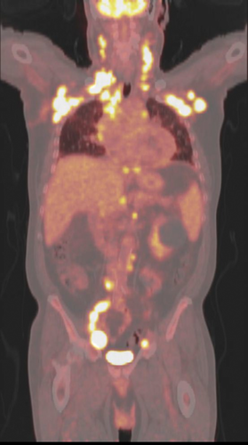
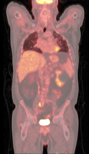
A 69-year-old male with rheumatoid arthritis, on methotrexate and plaquenil, presented with palpable right axillary lymphadenopathy. This was associated with progressive dyspnea, weight loss, and night-sweats for one-month duration. On examination, there was a 2 × 2 cm nontender lymph node on the right axillary. Positron emission tomography-computed tomography (PET-CT) illustrated diffuse hypermetabolic lymphadenopathy with moderate pericardial effusion (Image 1). Patient had a right axillary excisional biopsy that confirmed Diffuse Large B-Cell Lymphoma, which positively stained for Epstein–Barr virus encoded RNA (EBER) on in-situ-hybridization. Bone marrow biopsy was normal. A diagnosis of IIIB other iatrogenic immunodeficiency associated lymphoproliferative disorder (OIIA-LPD) was given and decided to hold methotrexate. After 6 weeks of withdrawal, the symptoms resolved along with his lymphadenopathy on PET-CT (Image 2). Patients with rheumatoid arthritis have a risk of developing LPDs with or without methotrexate. However, further immunosuppression with methotrexate and reactivation of Epstein–Barr virus is postulated to be a possible culprit in lymphomagenesis, given that withdrawal of methotrexate correlates with regression of disease in many cases 1-7. Patient currently remains in complete remission.
Details are in the caption following the image
PET-CT showing diffuse hypermetabolic uptake while on methotrexate.
Details are in the caption following the image
PET-CT illustrating no hypermetabolic activity after methotrexate withdrawal.

    The full text of this article hosted at iucr.org is unavailable due to technical difficulties.