Volume 11, Issue 6 pp. 1648-1649
interACTN Case
Open Access

The case of an 85-year-old woman with subacute onset of bilateral chorea

Nathan J. Nakatsuka

Nathan J. Nakatsuka

Harvard Medical School, Boston, Massachusetts, USA

Search for more papers by this author
Vihang Nakhate

Vihang Nakhate

Harvard Medical School, Boston, Massachusetts, USA

Department of Neurology, Brigham and Women's Hospital, Boston, Massachusetts, USA

Department of Neurology, Massachusetts General Hospital, Boston, Massachusetts, USA

Search for more papers by this author
Daniel S. Harrison

Daniel S. Harrison

Harvard Medical School, Boston, Massachusetts, USA

Department of Neurology, Brigham and Women's Hospital, Boston, Massachusetts, USA

Department of Neurology, Massachusetts General Hospital, Boston, Massachusetts, USA

Search for more papers by this author
Kristin M. Galetta

Kristin M. Galetta

Harvard Medical School, Boston, Massachusetts, USA

Department of Neurology, Brigham and Women's Hospital, Boston, Massachusetts, USA

Search for more papers by this author
Abby L. Olsen

Corresponding Author

Abby L. Olsen

Department of Neurology, University of Pittsburgh, Pittsburgh, Pennsylvania, USA

Department of Neurology, UPMC, Pittsburgh, Pennsylvania, USA

Search for more papers by this author
First published: 03 May 2024

Summary of Case (HPI, Relevant Examination Findings, and Relevant Data)

An elderly woman presented with subacute onset of chorea following a hospitalization for severe, uncontrolled diabetes, a urinary tract infection, and 6 months of depression. Her neurological examination demonstrated bilateral chorea involving the arms, legs, and jaw. MRI of the brain demonstrated bilateral T1 hyperintensity. She was diagnosed with diabetic striatopathy, also known as nonketotic hyperglycemic chorea, a rare complication of diabetes mellitus that classically causes hemichorea in the setting of very high blood glucose without ketosis. This case demonstrates typical imaging findings of diabetic striatopathy despite several atypical clinical features, including bilateral chorea, development of symptoms weeks after improvement in blood glucose, and demographics of the patient.

Diagnosis

Diabetic striatopathy.1, 2

Take-Home Points

  • Diabetic striatopathy, or nonketotic hyperglycemia induced chorea, occurs in the setting of markedly elevated blood glucose.
  • The mainstay of treatment is lowering of blood glucose, though, as our case indicates, chorea can start after this occurs.
  • If normalization of glucose does not resolve symptoms, additional treatments include neuroleptics and benzodiazepines.
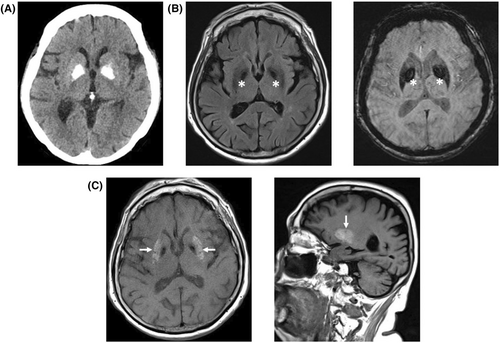
  • The typical imaging finding of diabetic striatopathy is T1 hyperintensity in the putamen (Fig. 1).
Details are in the caption following the image
CT head and MRI brain showing globus pallidus calcifications and putaminal T1 hyperintensity. (A) Non-contrast head CT axial image showing bilateral hyperdensities in the globus pallidi consistent with age-related calcification, unchanged from CT head 1 year prior (not shown). (B) MRI brain axial T2 FLAIR sequence (left) showing T2 hypointensities in bilateral globus pallidi (indicated by white asterisk) corresponding to hyperdensities on CT; SWI sequence (right) showing susceptibility artifact in bilateral globus pallidi (indicated by white asterisk) corresponding to CT hyperdensities, consistent with age-related calcification. (C) MRI brain axial (left) and sagittal (right) T1 pre-contrast sequences showing hyperintensity of bilateral putamen (indicated by white arrows).

    The full text of this article hosted at iucr.org is unavailable due to technical difficulties.