Developmental regression and mitochondrial function in children with autism
Funding Information
No funding information provided.
Abstract
Background
Developmental regression (DR) occurs in about one-third of children with Autism Spectrum Disorder (ASD) yet it is poorly understood. Current evidence suggests that mitochondrial function in not normal in many children with ASD. However, the relationship between mitochondrial function and DR has not been well-studied in ASD.
Methods
This cross-sectional study of 32 children, 2 to 8 years old with ASD, with (n = 11) and without (n = 12) DR, and non-ASD controls (n = 9) compared mitochondrial respiration and mtDNA damage and copy number between groups and their relation to standardized measures of ASD severity.
Results
Individuals with ASD demonstrated lower ND1, ND4, and CYTB copy number (Ps < 0.01) as compared to controls. Children with ASD and DR had higher maximal oxygen consumption rate (Ps < 0.02), maximal respiratory capacity (P < 0.05), and reserve capacity (P = 0.01) than those with ASD without DR. Coupling Efficiency and Maximal Respiratory Capacity were associated with disruptive behaviors but these relationships were different for those with and without DR. Higher ND1 copy number was associated with better behavior.
Conclusions
This study suggests that individuals with ASD and DR may represent a unique metabolic endophenotype with distinct abnormalities in respiratory function that may put their mitochondria in a state of vulnerability. This may allow physiological stress to trigger mitochondrial decompensation as is seen clinically as DR. Since mitochondrial function was found to be related to ASD symptoms, the mitochondria could be a potential target for novel therapeutics. Additionally, identifying those with vulnerable mitochondrial before DR could result in prevention of ASD.
Introduction
Autism Spectrum Disorder (ASD) affects 1 in 59 children.1 Despite intensive research, the etiologies of ASD are still unclear. One area of promising research is the link between mitochondrial function and ASD. Mitochondrial function is important in tissues with high-energy demand, notably the brain, gastrointestinal tract, and immune system – all systems which are commonly dysfunctional in individuals with ASD.2, 3
An important distinction when discussing abnormalities of mitochondrial physiology is the difference between mitochondrial disease and mitochondrial dysfunction. Meta-analysis has demonstrated that 5% of children with ASD can be diagnosed with the very circumscribed diagnosis of mitochondrial disease, which is defined by very low electron transport chain (ETC) complex activity or a clear genetic defect.4 In contrast, the same meta-analysis found that 30%-50% of individuals with ASD demonstrate biomarkers of mitochondrial dysfunction.4, 5 Other studies suggest that up to 80% of individuals with ASD show abnormal ETC activity in lymphocytes and granulocytes6, 7 as well as postmortem brain.8 Thus, it is hypothesized that individual with ASD have unique changes in mitochondrial function that may be distinct from classically defined mitochondrial disease.4, 9 For example, rather than significant decreases is ETC complex activity, individuals with ASD have been found to have significant elevations in ETC complex activity in multiple tissues.8, 10-14 In a model of mitochondrial dysfunction in ASD, a subset of lymphoblastoid cell lines (LCLs) from boys with ASD have been shown to have elevated respiratory rates and found to be sensitive to physiological stress.15, 16
About 32% of children with ASD undergo an enigmatic developmental regression (DR) during the second year of life.17, 18 Following apparently normal development, DR involves loss of the child’s previously acquired milestones followed by the emergence of ASD symptoms.17, 19-23 DR most often appears clinically after childhood illness, often with fever.24, 25 Atypical DR (e.g., multiple regressions)26 and DR accompanied by fever25 has been associated with mitochondrial disease in ASD. Despite this association, most studies studying mitochondria function in relation to ASD have not closely examined DR.
Our goal was to study the relationship between DR and mitochondrial bioenergetics in children with ASD. We hypothesized that DR is triggered in a subset of children with ASD due to underlying abnormalities in mitochondrial function. We hypothesize that children with ASD and DR have marginal but adequate mitochondrial function until a trigger increases physiological stress that overwhelms the capacity of mitochondria to function, resulting in clinically observed DR. Marginal mitochondrial function could be represented by very low mitochondrial activity, as seen in mitochondrial disease, but we believe that marginal mitochondrial function could also be represented by very high mitochondrial activity, as seen in the LCL model since such elevated respiratory rates could represent a state in which the mitochondria is close to its maximum ability to function, making mitochondria sensitivity to physiological stress.9, 15, 16 To this end, this study examined mitochondrial function and mitochondrial DNA (mtDNA) damage and copy number in peripheral blood mononuclear cells (PBMCs) from three age-matched groups of children: those with ASD, with and without DR, and non-ASD controls.
Materials and Methods
Participants
The protocol was approved by the University of Massachusetts Medical Center, Worcester, Institutional Review Board. Following informed written consent of the parents and assent by children who were able, 10–15cc of blood was drawn by venipuncture. The diagnosis of ASD and history of DR were confirmed by a single clinician (AWZ) from extensive medical history, examination and review of medical records. Symptoms of ASD were measured for each child with ASD using the clinician-rated Ohio Autism Clinical Impression Scale–Severity (OACIS-S)27 and parent-completed Aberrant Behavior Checklist (ABC)28 and Social Responsiveness Scale-2 (SRS).29
Bioenergetic measurements
PBMCs were isolated within 2 h of phlebotomy using Histopaque 1077 protocol. PBMCs were counted, suspended in culture medium (90% FBS/10% DMSO) and frozen at −80°C in a specialized Mr Frosty cryofreeze container (Thermo Fisher Scientific, Waltham, MA). PBMCs were transferred to LN3 liquid nitrogen chamber within 24 h and transferred to the University of Arizona laboratory for analysis on dry ice by overnight carrier within 12 months of collection. Upon receipt, they were stored in LN3 liquid nitrogen chamber until Seahorse assay. PBMC average (standard deviation; range) viability was 48% (16%, 22%–65%), 57% (12%; 26%–80%), and 44% (19%, 27%–70) for ASD/No-DR, ASD/DR, and CNT groups, respectively, and was not significantly different between groups. In our experience, a viability of 20% or greater provides reliable Seahorse measurements.
PBMC’s were resuspended in RPMI medium (Life technologies, Gibco, Grand Island, NY) supplemented with 1 mmol/L pyruvate, 2 mmol/L glutamax, and 25 mmol/L glucose, warmed to 37°C and pH adjusted to 7.4 prior to cell suspension. XFe96 plates (Seahorse Bioscience, Billerica, MA) were prepared by adding 25 μL of 50 μg/mL Poly-d-lysine (EMD Millipore, Billerica, MA) for 2 h, washing with 250 μL sterile water and drying in a laminar flow hood overnight prior to seeding with 600 k viable PBMCs per well. All available PMBCs were utilized resulting in 1–8 wells per participant, thus allowing for several replicates per participant. One ASD/DR and two CNT participants only had one well, so their data were not used. However, inclusion of their data did not change any results significantly.
After seeding, the plates were spun with slow acceleration (4 on a scale of 9) to a maximum of 100 g for 2 min and then allowed to stop with zero braking (Eppendorf Model 5810R Centrifuge). The plate orientation was reversed, and the plate was spun again to 100 g in the same fashion. Prior to Seahorse assay, XFe96 wells were visualized using an inverted microscope to ensure that PBMCs were evenly distributed in a single layer and viability of the cells was confirmed by trypan blue exclusion.
Mitochondrial bioenergetics were assessed using the Seahorse XFe96 extracellular flux analyzer by measuring oxygen consumption rate (OCR) and deriving bioenergetic measures. OCR is a fundamental measure of mitochondrial function and reflects coupled mitochondrial respiration as well as uncoupled consumption of oxygen which is used to regulate reactive oxygen species at the inner mitochondrial membrane.30-33 Figure 1 depicts the typical Seahorse flux analyzer assay and its various parameters.
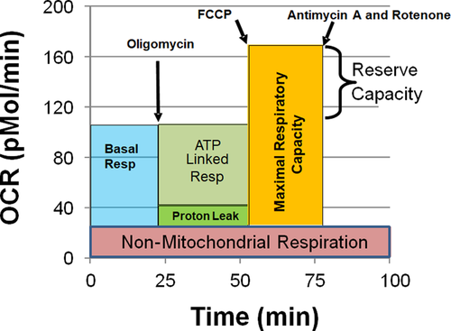
- Baseline OCR: Baseline OCR is measured before introducing any reagents.
- OCR after oligomycin: Oligomycin, a complex V inhibitor, shuts down the production of ATP so the OCR related to ATP production can be determined.
- Maximal OCR: Carbonyl cyanide-p-trifluoromethoxyphenyl-hydrazone is used to collapse the mitochondrial inner membrane gradient, inducing the mitochondria to function at its maximum extent possible.
- Nonmitochondrial OCR: Antimycin A and Rotenone are added to shut down ETC complex activity to measure OCR from non-ETC processes.
- Extracellular acidification rate = Rate of free protons production in the microchamber at baseline conditions.
- ATP-linked respiration: The fraction of basal OCR that is attributed to ATP production

- Proton-leak respiration: The amount of basal OCR that is associated with protons leaking through the inner mitochondrial membrane in order to control oxidative stress

- Maximal respiratory capacity: The maximum respiratory rate of the ETC. This parameter is thought to be sensitive to deficits in mitochondrial biogenesis, mtDNA damage and/or inhibition of ETC function.

- Reserve capacity: The amount of extra ATP that can be produced by oxidative phosphorylation when there is a sudden increase in energy demand. This parameter is an index of mitochondrial health; when it becomes negative, the mitochondrion is unhealthy leading toward apoptosis.

- Coupling efficiency: The amount of ATP made per atom of oxygen consumed. This parameter provides a measure of how much of the mitochondria’s total respiratory rate is contributing to the production of energy and how much is wasted to control oxidative stress.

- Bioenergetic health index (BHI): The BHI combines multiple measures for mitochondrial respiration into one index which represents overall mitochondrial function and health.34 The index represents the mitochondrial energy production corrected for oxygen consumption by sources which do not produce energy. The BHI has been developed using the Seahorse platform to measure mitochondrial function in peripheral immune cells34 and is proposed as a biomarker of bioenergetic health.35-38

- Proton efflux rate: A measure of proton flux corrected for important assay factors. Since Proton Efflux Rate is a linear transformation of the Extracellular Acidification Rate, only the Proton Efflux Rate is analyzed.

Derived measures were calculated for each well. Measurements were averaged across wells for each participant to obtain one value per participant. Wells with nonphysiological ATP-Linked Respiration and Proton-Leak Respiration values (<−2 pMol/min) were not used.
Mitochondrial DNA (mtDNA) analysis
Genomic DNA was isolated from PBMCs from 11 ASD (7 ASD/DR, 4 ASD/No-DR) and 7 CNT participants using QIAamp DNA Mini Kit (QIAGEN, Germantown, MD). The DNA concentration was measured by the spectrophotometer Nano Drop 2000C (Thermo Scientific). Toyobo Thunderbird™ SYBR qPCR master mix was used with standard block protocol in BIO-RAD CFX96 Real-Time System.
The human RT-PCR mtDNA damage analysis kit (Detroit R&D, Inc.) measures the amount of damage to long (8.1–8.8 kb) mtDNA pieces. A higher product represents less mtDNA damage. The PCR DNA volume (2 µL) product from the qPCR was optimized by serially diluting (10-to-10,000 fold) with the nuclease free water. The quantification of the RT-PCR products was calculated against the standard curve using threshold cycle (Ct) value and the DNA concentration (log scale) of each of the 8.8 kb mtDNA standards. Samples were run in duplicate and averaged.
The NovaQUANT™ Human Mitochondrial-to-Nuclear DNA ratio Kit (EMD Millipore Corporation) measured quantities of two nuclear (BECN and NEB) and two mitochondrial (ND1 and ND6) genes. Positive (human wt) and negative (rho zero) controls were included for mitochondrial targets. A ‘no template control’ NTC using the DNase/RNase free water was also used. The abundance of each gene was estimated from the corresponding cycle threshold (Ct) value. The Ct differences (∆Ct) between ND1/BECN and ND6/NEB was used to estimate copy number (2∆Ct). The average ∆Ct between BECN and NEB was 0.34 (SD = 0.24) which suggests technical accuracy (<1.0).
Copy number of NADH Dehydrogenase 4 (ND4) and Cytochrome B (CYTB) genes were also measured using the pyruvate kinase (PK) gene as the nuclear gene reference. DNA primer sequences reported by Gu et al.39 were synthesized by Integrated DNA Technologies Inc (Coralville, IA). A Toyobo Thunderbird™ SYBR qPCR master mix (Toyobo Life Sciences, Japan) with standard block protocol in BIO-RAD CFX96 Real Time System (Bio-Rad Laboratory Inc, Hercules, CA) was used to denature at 95°C for 10 min followed by 40 cycles at 95°C for 15 sec and 60°C for 1 min. Each 20 µL RT-PCR reaction mixture contained 9ng of isolated genomic DNA, 0.4 µmol/L forward primers 0.4 µmol/L reverse primers, and 10 µL 2xSYBR qPCR master mix. A ‘no template control’ NTC using the DNase/RNase free water was also included. The Ct differences (∆Ct) between ND4/PK and CYTB/PK were used to calculate copy number (2∆Ct).
Statistical analysis
Two one-way Analysis of Variance (ANOVA) were used to compare OCR rates, derived parameters, and mtDNA measurements across groups (ASD vs. CNT and ASD/DR vs. ASD/No-DR) using SAS (Version 14.1, SAS Institute, Cary, NC). Linear regression models (SAS procedure glimmix) was used to analyze the relationship between behavioral measures and derived bioenergetic and mtDNA parameters including only ASD participants. The regression models included the factor of DR and its interaction. Because of the large number of behavioral measures (23 measures), the Bonferroni correction was used to set the P-value cut-off (0.05/23 = 0.002). To better understand relationships between mitochondrial measurements and behavior in regression models which demonstrated significant DR interactions, correlations were performed between the behavioral measures and the derived parameters for the DR groups separately.
Results
Participant characteristics
Table 1 and Table S1 provide participant characteristics. There were no statistically significant differences between the CNT, ASD/DR, and ASD/No-DR groups. There were few small but significant differences between behavioral measures across ASD groups.
| Variable | ASD/DR | ASD/No-DR | Controls | P-value |
|---|---|---|---|---|
| Number of participants | 11 | 12 | 9 | – |
| Gender (female) | 1/11 (9%) | 3/12 (25%) | 4/9 (44%) | |
| Age (years) ± SD | 7.13 ± 1.7 | 5.14 ± 1.5 | 6.60 ± 2.1 | 0.06 |
| OACIS-S scores ± SD | ||||
| General level of autism | 4.8 ± 0.9 | 5.3 ± 0.9 | – | 0.20 |
| Social interaction | 4.8 ± 0.9 | 5.5 ± 0.8 | – | 0.07 |
| Aberrant behaviors | 4.5 ± 0.9 | 4.8 ± 0.9 | – | 0.40 |
| Repetitive behaviors | 4.5 ± 1.6 | 4.6 ± 1.1 | – | 0.80 |
| Verbal communication | 4.8 ± 1.1 | 5.6 ± 1.2 | – | 0.08 |
| Non-verbal communication | 4.4 ± 0.8 | 5.2 ± 0.8 | – | 0.02 * |
| Hyperactivity/inattention | 4.6 ± 1.1 | 5.2 ± 1.0 | – | 0.20 |
| Anxiety | 4.2 ± 1.6 | 3.9 ± 1.1 | – | 0.70 |
| Sensory sensitivities | 4.2 ± 0.8 | 4.9 ± 0.8 | – | 0.04 * |
| Restricted/narrow interests | 4.2 ± 1.6 | 4.5 ± 0.7 | – | 0.50 |
| Aberrant behavior checklist ± SD | ||||
| Total score | 48.6 ± 24.7 | 65.1 ± 25.3 | – | 0.10 |
| Subscale irritability | 11.8 ± 6.6 | 12.8 ± 7.4 | – | 0.80 |
| Subscale lethargy | 8.8 ± 6.9 | 14.2 ± 5.9 | – | 0.06 |
| Subscale stereotypy | 7.4 ± 4.7 | 9.1 ± 5.6 | – | 0.50 |
| Subscale hyperactivity | 16.1 ± 10.5 | 25.2 ± 9.6 | – | 0.04 * |
| Subscale inappropriate speech | 4.5 ± 2.5 | 3.8 ± 3.0 | – | 0.60 |
| Social responsiveness scale ± SD | ||||
| Total raw score | 106.8 ± 21.6 | 111.7 ± 23.2 | – | 0.60 |
| Awareness raw score | 14.1 ± 2.9 | 13.4 ± 2.9 | – | 0.50 |
| Cognition raw score | 20.2 ± 3.6 | 21.2 ± 5.1 | – | 0.60 |
| Communication raw score | 36.8 ± 9.0 | 37 ± 9.5 | – | 0.90 |
| Motor skills raw score | 15.3 ± 5 | 18.9 ± 3.6 | – | 0.08 |
| Repetitive behaviors raw score | 20.3 ± 5.9 | 21.4 ± 5.8 | – | 0.70 |
| SCI raw score | 86.5 ± 17.7 | 90.3 ± 18.1 | – | 0.60 |
- * Significant P-values are shown in bold.
Mitochondrial respiration
- At 59.8 min, the ASD/DR group (145.8 ± 70.1) had 78% higher OCR than the ASD/no-DR group (81.8 ± 49.9) and 118% higher OCR than CNT group (66.8 ± 56.4).
- At 66.3 min, the ASD/DR group (111.7 ± 54.2) had 78% higher OCR than the ASD/no-DR group (62.7 ± 37.7) and 103% higher OCR than CNT group (55.0 ± 44.8).
- At 72.8 min, the ASD/DR group (93.5 ± 46.5) had 75% higher OCR than the ASD/no-DR group (53.5 ± 31.9) and 97% higher OCR than CNT group (47.5 ± 39.4).
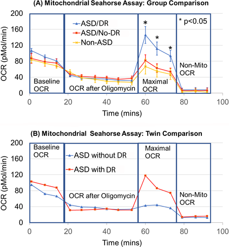
To examine this in more detail, a pair of 7-year-old male fraternal twins, both diagnosed with ASD but only one with DR were examined in more detail. As seen in Figure 2B, Maximal OCR is obviously much higher in Twin A with ASD/DR compared to Twin B with ASD/No-DR.
Respiratory parameters
Figure 3 demonstrates the derived respiratory parameters separately for the clinical group (ASD vs. CNT) and regression status (DR vs. No-DR). One of the most obvious findings is the variability in respiratory parameters for the ASD groups as compared to the control group. There was no significant difference between the average ASD and CNT values.
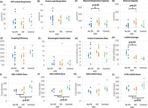
The two groups of participants with ASD demonstrated differences in respiratory parameters depending on whether nor not they had a history of DR. Those with DR demonstrated significantly higher Maximal Respiratory Capacity [F(1,21) = 4.71, P < 0.05; Fig. 3C] and Reserve Capacity [F(1,21) = 7.41, P = 0.01; Fig. 3D].
mtDNA analysis
Overall mtDNA damage was not significantly different between ASD and CNT participants but was lower (higher amount of 8.8kb mtDNA segments) in ASD/No-DR participants compared to ASD/DR participants [F(1,9) = 6.40, P < 0.05] (Fig. 3H). The relative number of ND1 [F(1,17) = 9.86, P < 0.01; Fig. 3I], ND4 [F(1,17) = 13.72, P < 0.01; Fig. 3J] and CYTB [F(1,17) = 8.64, P < 0.01; Fig. 3L] gene copies was significantly reduced in ASD participants as compared to the CNT participants. The copy number of ND6 (Fig. 3L), the only gene tested which resides on the mtDNA light-chain, was not different with respect to participant group or DR status.
Associations between behavioral measures and mitochondrial function
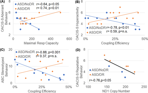
As seen in Figure 4A–C the relationship between behavior and derived respiratory parameters was different depending on the history of DR for the OACIS-S subscales of Aberrant Behavior and Maximal Respiratory Capacity [F(1,16) = 14.85, P = 0.001; Fig. 4A] and Hyperactivity and Coupling Efficiency [F(1,16) = 17.69, P < 0.001; Fig. 4B] and for the ABC Stereotyped Behavior subscale and Coupling Efficiency [F(1,16) = 17.42, P < 0.001; Fig. 4C]. In general, for those without a history of DR higher Maximal Respiratory Capacity or Coupling Efficiency was associated with less severe behaviors while for those with a history of DR higher Maximal Respiratory Capacity or Coupling Efficiency was associated with more severe behaviors. In general the relationships between behavior and mitochondrial respiration were stronger for the ASD participants without a history of DR. Higher ND1 copy number was associated with better (i.e., lower) OACIS-S subscales of Repetitive Behavior [F(1,5) = 45.66, P = 0.001] (Fig. 4D).
Discussion
DR is an important feature that affects about one-third of children with ASD.17 To better understand the significance of mitochondrial function in DR, we examined bioenergetics using the Seahorse XF96 analyzer in live immune cells derived from controls and children with ASD, both with and without DR. We have previously documented abnormal mitochondrial function in PBMCs using the Seahorse XF96 in siblings with ASD and genetic abnormalities40 and in children with ASD and immune abnormalities.41 Our data demonstrate that differences in mitochondrial respiration are found in children with ASD depending on a history of DR, and that mitochondrial respiration and mtDNA ND1 copy number are associated with ASD symptoms, particularly in those without a history of DR. This suggests that variations in mitochondrial function are important in understanding the underlying physiological abnormalities in children with ASD, especially regarding their clinical characteristics.
Relationship between ASD and mitochondrial DNA
Data from this study suggests that young children with ASD, regardless of whether they demonstrated DR or not, have significantly lower copy number of important mtDNA genes associated with ETC function, including ND1 and CYTB. A large study (n = 122) of leukocytes in individuals with ASD and intellectual disability found a decrease in ND1 copy number associated with ASD.42 While a small study (n = 14) of ASD brain tissue found a relative increase in ND1 copy number,39 the same study found deletions in the same genes and decreased ETC activity.39 Additionally, other studies have found deletions in CYTB6, 7 which is another gene found to have a decreased copy number in this study. Higher ND1 copy number was related to less aberrant behavior. Since this study found a reduction in ND1 copy number relative to CNT, this relationship could suggest more normal ND1 copy number, and thus possibly more normal ETC Complex 1 function, could be related to less disruptive behavior.
ASD/No-DR participants demonstrated less mtDNA damage compared to ASD/DR. Although mtDNA damage has not been examined in ASD in the past, other studies have noted an increased mtDNA copy number in leukocytes6, 7, 43-45 and buccal mucosa.46 An increase in overall mtDNA content is believed to be a compensatory reaction that maintains adequate wild-type mtDNA in the face of high levels of oxidative stress which itself can damage and cause deletions in mtDNA.7 Thus, it is possible that these data suggest that ASD/No-DR individuals are better able to regulate oxidative stress, thereby reducing mtDNA damage.
Relationship between developmental regression and mitochondrial function
The ASD/DR group demonstrated higher respiration rates as compared to the ASD/No-DR group. First, the maximal OCR of the ASD/DR group was nearly double that of the ASD/No-DR group. In a pair of fraternal twins, the twin with DR had more than twice the peak OCR compared to his twin without DR. Second, Maximal Respiratory Capacity and Reserve Capacity were higher in the ASD/DR group as compared to ASD/No-DR group.
Rose et al.16 hypothesized that higher than normal Reserve Capacity may be a protective adaptation in response to environmental stressors while Bennuri et al47 showed that prolonged exposure to low-levels of oxidative stress could induce this increase in baseline Reserve Capacity. However, this increase in Reserve Capacity is associated with a sensitivity to oxidative stress such that lower levels of reactive oxygen species are needed to deplete Reserve Capacity, leading to mitochondrial dysfunction. Thus, we believe that the subset of LCLs with increased Reserve Capacity are a good model for DR.
Previous reports have linked mitochondrial disease in children with25, 26, 48 and without49-51 ASD to DR, especially with an associated trigger. This is the first study to provide evidence for a unique type of mitochondrial dysfunction, that is most likely distinct from mitochondrial disease, to be related to ASD with DR. Thus, children with ASD and DR make up an important subset of children with ASD that require further careful study of their mitochondria. Interestingly, previous in vitro studies have shown that N-acetylcysteine (NAC) protects LCLs with high respiratory rates from losing Reserve Capacity under physiological stress,52 suggesting a potential prophylactic treatment for patients with vulnerable mitochondria to prevent developmental regression and possibly prevent ASD or at least mitigate symptom severity.
Limitations
Our study is not without limitations. We have a small sample size due to the difficulty in confirming DR and age matching to children with ASD without DR and controls. Indeed, our novel findings need to be validated in a larger sample. The BHI needs to be studied in a larger sample where the relative weights of different components of the BHI can be examined. Nevertheless, our data provide evidence that our patients with ASD and DR may have impaired mitochondrial function.
Conclusions
In this study we found that specific parameters of mitochondrial function vary with ASD symptomatology including behavior and history of developmental regression. This suggests that mitochondria have a direct effect on the severity of ASD symptoms and opens the possibility that interventions targeting the mitochondria could have a role in the prevention of developmental regression and treating ASD symptomatology. Additionally, these important clinical factors, especially developmental regression, needs to be considered when recruiting individuals with ASD to study mitochondrial function as the participant characteristics will directly affect the mitochondrial function of the sample.
Acknowledgments
We thank the children, their parents and families who participated in the study, for their interest and commitment. Patients were referred to the study by Roula Choueiri, JoAnn Carson, Stephanie Blenner and others in the Department of Pediatrics at UMass Memorial Children’s Medical Center. The nursing staff of the UMass Memorial Pediatric Outpatient Department provided valuable assistance with phlebotomies.
Author Contributions
K.S,, I.N.S A.W.Z., and R.E.F contributions to the conception and design of the work and data analysis and interpretation. E.D. and S.L.C. contributed to the acquisition of the data and biological samples. I.N.S., M.K., and D.L contributed to the analysis of biological samples. K.S, I.N.S, A.W.Z., M.K., and R.E.F drafted the initial manuscript work. All authors revised the manuscript. Each author has approved the submitted version of the manuscript and agrees both to be personally accountable for their own contributions and to ensure that questions related to the accuracy or integrity of any part of the work, even ones in which the author was not personally involved, are appropriately investigated, resolved, and the resolution documented in the literature.
Conflict of Interest
The authors declare no conflicts of interest.


