Standard Article

Assembly of a Functional Triiron(III) Cluster in L-Ferritin

Silvia Ciambellotti

Silvia Ciambellotti

Magnetic Resonance Center (CERM), University of Florence, Florence, Italy

Department of Chemistry “Ugo Schiff”, University of Florence, Florence, Italy

Search for more papers by this author
Cecilia Pozzi

Cecilia Pozzi

Department of Biotechnology, Chemistry and Pharmacy, University of Siena, Siena, Italy

Search for more papers by this author
Stefano Mangani

Stefano Mangani

Magnetic Resonance Center (CERM), University of Florence, Florence, Italy

Department of Biotechnology, Chemistry and Pharmacy, University of Siena, Siena, Italy

Search for more papers by this author
Paola Turano

Paola Turano

Magnetic Resonance Center (CERM), University of Florence, Florence, Italy

Department of Chemistry “Ugo Schiff”, University of Florence, Florence, Italy

Search for more papers by this author

Abstract

The L-subunits of mammalian ferritins are generally accepted to facilitate iron biomineral formation by providing nucleation sites. The formation of a μ3-oxo trinuclear iron cluster on the inner cage of homopolymeric recombinant human L ferritin and natural horse spleen ferritin has been observed upon diffusion of ferrous ions through metal-free ferritin crystals. Three glutamate side chains (Glu60, Glu61, and Glu64) act as bridging ligands between iron pairs, thus driving the cluster assembly. In the fully formed cluster, observed after 60′ diffusion in the human L ferritin, the iron ions are also bridged by peroxide anions, which could originate from ferrous iron oxidation by dioxygen. Substitution of Glu60, Glu61, and Glu64 by alanine residues significantly reduces the iron biomineralization rates, thus suggesting that the observed cluster represents the biomineral nucleation site.

3D Structure
Description unavailable

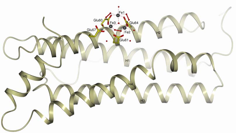
Ribbon representation of the 3D structure of a single subunit of iron-loaded HuLf (PDB code: 5lg8). The protein anchors a triiron cluster on the subunit surface facing the inner cage. The side chain residues of Glu60, Glu61, Glu64, and of the shuttling residue Glu57 are shown as sticks. Iron(III) atoms are depicted as gray spheres. All the structural pictures have been generated using the molecular-graphic software CCP4mg.1

The full text of this article hosted at iucr.org is unavailable due to technical difficulties.