5-Aminolaevulinic Acid Dehydratase
Abstract
The enzyme 5-aminolaevulinic acid dehydratase (ALAD, E.C.4.2.1.24), sometimes referred to as porphobilinogen synthase, catalyzes the second step in the biosynthesis of tetrapyrroles involving the condensation between two 5-aminolaevulinic acid (ALA) molecules to form the pyrrole porphobilinogen (PBG). The X-ray structures of several ALADs have been determined showing that the enzyme forms a large homo-octameric structure with all eight active sites on the outer surface. Each subunit adopts the triose phosphate isomerase (TIM) barrel fold with an N-terminal arm, which forms extensive intersubunit interactions. Pairs of monomers associate with their arms wrapped around each other to form compact dimers. Four dimers, which interact principally via their arm regions, form the octamer, which has a large solvent-filled cavity in the center. The active site of each subunit is located in a pronounced cavity formed by loops at the C-terminal ends of the strands forming the TIM barrel.
In humans, hereditary deficiencies in ALAD give rise to the rare disease Doss porphyria, and the sensitivity of the enzyme to inhibition by lead ions is thought to be one of the main problems in acute lead poisoning. ALADs from many species (except plants and some bacteria) have a zinc ion at the active site, which has been implicated in the catalytic mechanism and is readily displaced by the inhibitor lead. These ALADs have a cysteine-rich motif that coordinates the active site zinc ion (α-site). In contrast, ALADs from plants and some bacteria have a number of mutations in the metal-binding motif that replace the cysteine residues with other amino acids. Two of the cysteines are replaced by aspartate, which is thought to be more appropriate for coordination of magnesium ions, which these enzymes apparently require instead of zinc. Some ALADs have an additional zinc or magnesium binding site located between the subunits of the octamer (β-site) that has more of a structural or activating role.
Early inhibitor binding studies defined the interactions made by only one of the two substrate moieties (P-side substrate) that bind to the enzyme during catalysis. Biochemical and crystallographic data showed that P-side substrate binds by forming a Schiff base with the enzyme at Lys247 (Escherichia coli ALAD numbering). Until very recently there has been little evidence that binding of the second substrate moiety (A-side substrate) involves formation of a Schiff base with the enzyme. However, the structures of diacid inhibitors provided an improved definition of the interactions made by both the substrate molecules (A- and P-side). The most intriguing result was the finding that 4,7-dioxosebacic acid forms a second Schiff base with the enzyme involving Lys195 (E. coli ALAD numbering), suggesting that A-side substrate makes the same interaction during catalysis. This finding has been substantiated by the demonstration that the inhibitor 5-fluorolaevulinic acid binds by making Schiff bases with both the active site lysine residues. Current proposals for the catalytic mechanism involve the binding of both substrate moieties by formation of Schiff bases with the active site lysines.
3D Structure
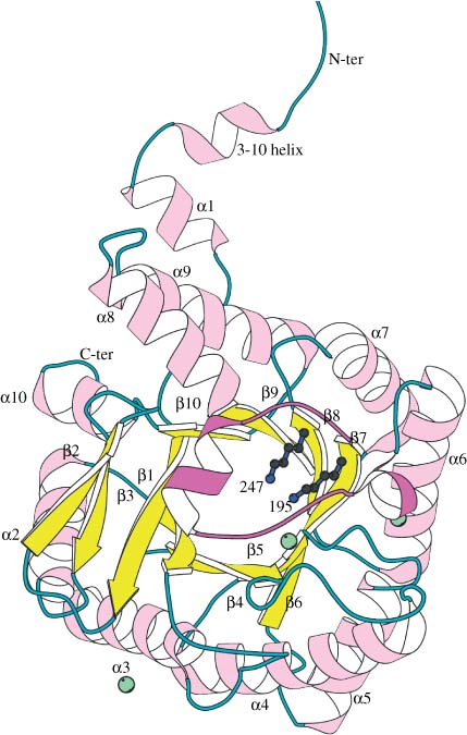
Shows a ribbon diagram of the fold of the E. coli ALAD monomer (PDB code 1B4E). The dominant feature of the tertiary structure is the closed (α/β)8 or TIM-barrel with the active site located in a pronounced cavity (facing reader). Helical segments are shown in lilac, strands in yellow, and the connecting loop regions in cyan. The active site flap (197–220) is colored magenta and the enzyme's N-terminal arm region (residues 1–30), which forms extensive quaternary contacts, is shown above the barrel. The two adjacent lysines implicated in catalysis (195 and 247) are shown in blue. Lys247 forms a Schiff base link with P-site ALA. Green spheres indicate the locations of the zinc binding sites. The active site zinc ion held by three cysteines can be seen close to the two lysines in the center of the TIM-barrel. The zinc ion on the right-hand side (β-site) is involved in intersubunit contacts and that on the lower left is involved in crystal contacts. This figure was prepared using the program BOBSCRIPT.30


