X-Ray Photoelectron Spectroscopy of Porphycenes: Charge Asymmetry Across Low-Barrier Hydrogen Bonds
Abhik Ghosh Prof. Dr.
Institute of Chemistry University of Tromsø 9037 Tromsø (Norway) Fax: (+47) 77644765
Search for more papers by this authorJohn Moulder Dr.
Physical Electronics, Inc. 6509 Flying Cloud Drive, Eden Prairie, MN 55344 (USA)
Search for more papers by this authorMartin Bröring Dr.
Institut für Anorganische Chemie, Universität Würzburg Am Hubland, 97074 Würzburg (Germany)
Search for more papers by this authorEmanuel Vogel Prof. Dr.
Institut für Organische Chemie der Universität Greinstrasse 4, 50939 Köln (Germany)
Search for more papers by this authorAbhik Ghosh Prof. Dr.
Institute of Chemistry University of Tromsø 9037 Tromsø (Norway) Fax: (+47) 77644765
Search for more papers by this authorJohn Moulder Dr.
Physical Electronics, Inc. 6509 Flying Cloud Drive, Eden Prairie, MN 55344 (USA)
Search for more papers by this authorMartin Bröring Dr.
Institut für Anorganische Chemie, Universität Würzburg Am Hubland, 97074 Würzburg (Germany)
Search for more papers by this authorEmanuel Vogel Prof. Dr.
Institut für Organische Chemie der Universität Greinstrasse 4, 50939 Köln (Germany)
Search for more papers by this authorThis work was supported by the Norwegian Research Council, the VISTA program of Statoil (Norway), the Norwegian Academy of the Sciences and Letters, and the Deutsche Forschungsgemeinschaft. A.G. acknowledges stimulating discussions with Professor Thomas H. Morton.
Graphical Abstract
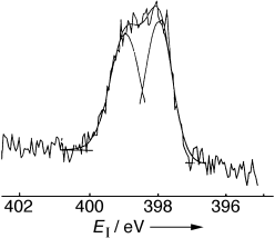
It is ironic that although it is typically a technique that “sees” all elements other than hydrogen atoms, X-ray photoelectron spectroscopy (XPS) acts as a sensitive probe of low-barrier hydrogen bonds, as seen in a study of free-base tetrapyrroles. The nitrogen 1s XPS peaks never quite coalesce, even when the N⋅⋅⋅H⋅⋅⋅N hydrogen bonds are almost perfectly symmetrical, as is the case for dibenzo[cde,mno]porphycenes (see picture).
References
- 1 W. W. Cleland, P. A. Frey, J. A. Gerlt, J. Biol. Chem. 1998, 273, 25 529.
- 2
- 2a B. Schwartz, D. G. Drueckhammer, J. Am. Chem. Soc. 1996, 118, 9826;
- 2b Y. Kato, L. M. Toledo, J. Rebek, Jr., J. Am. Chem. Soc. 1996, 118, 8575;
- 2c C. J. Smallwood, M. A. McAllister, J. Am. Chem. Soc. 1997, 119, 11 277.
- 3 B. Schiøtt, B. B. Iversen, G. K. H. Madsen, F. K. Larsen, T. C. Bruice, Proc. Natl. Acad. Sci. USA 1998, 95, 12 799;
- 3b B. Schiøtt, B. B. Iversen, G. K. H. Madsen, T. C. Bruice, J. Am. Chem. Soc. 1998, 120, 12 117.
- 4
- 4a G. A. Gerlt, P. G. Gassman, J. Am. Chem. Soc. 1993, 115, 11 552;
- 4b G. A. Gerlt, P. G. Gassman, Biochemistry 1993, 32, 11 943.
- 5 W. W. Cleland, M. M. Kreevoy, Science 1994, 264, 1887.
- 6 P. A. Frey, S. A. Whitt, J. B. Tobin, Science 1994, 264, 1927.
- 7 A. Warshel, A. Papazyan, Proc. Natl. Acad. Sci. USA 1996, 93, 13 665.
- 8
- 8a A. Warshel, A. Papazyan, P. A. Kollman, Science 1995, 269, 102;
- 8b W. W. Cleland, M. M. Kreevoy, Science 1995, 269, 104;
- 8c P. A. Frey, Science 1995, 269, 104.
- 9
- 9a E. Vogel, J. Heterocycl. Chem. 1996, 33, 1461;
- 9b for another account of the same area, see J. L. Sessler, S. J. Weghorn, Expanded, Contracted, and Isomeric Porphyrins, Elsevier, Amsterdam, 1997.
- 10
- 10a A. Ghosh in The Porphyrin Handbook, Vol. 7 ( ), Academic Press, New York, 2000, Chap. 47, p. 1;
- 10b A. Ghosh, Acc. Chem. Res. 1998, 31, 189.
- 11
- 11a A. Ghosh, J. Fitzgerald, P. G. Gassman, J. Almlöf, Inorg. Chem. 1994, 33, 6057;
- 11b A. Ghosh, J. Org. Chem. 1993, 58, 6932;
- 11c P. G. Gassman, A. Ghosh, J. Almlöf, J. Am. Chem. Soc. 1992, 114, 9990.
- 12 For other applications of XPS to the study of hydrogen bonds, see for example:
- 12a E. Hasselbach, A. Henriksson, F. Jachimowicz, J. Wirz, Helv. Chim. Acta 1972, 55, 1757;
- 12b K. Wozniak, H. Y. He, J. Klinowski, T. L. Barr, P. Milart, J. Phys. Chem. 1996, 100, 11 420.
- 13
- 13a T. Butenhoff, C. B. Moore, J. Am. Chem. Soc. 1988, 110, 8336;
- 13b Z. Smedarchina, M. Z. Zgierski, W. Siebrand, P. M. Kozlowski, J. Chem. Phys. 1998, 109, 1014;
- 13c for a review of NH tautomerism in porphyrins, see D. K. Maity, T. N. Truong, J. Porphyrins Phthalocyanines, in press.
- 14
- 14a C. B. Storm, Y. Teklu, J. Am. Chem. Soc. 1972, 94, 1745. For more detailed analyses of NH tautomerism in porphyrins, including isotope labeling, see
- 14b M. Schlabach, H.-H. Limbach, E. Bunnenberg, A. Y. L. Shu, B.-R. Tolf, C. Djerassi, J. Am. Chem. Soc. 1993, 115, 4554;
- 14c C. J. Medforth in The Porphyrin Handbook, Vol. 5 ( ), Academic Press, New York, 2000, Chap. 35, p. 1.
- 15 Among the free-base derivatives of tetrapyrrolic porphyrin isomers with N4 cores, porphycene is as stable as, or slightly more stable than, porphyrin. This reflects the presence of short, strong hydrogen bonds in free-base porphycenes: Y.-D. Wu, K. W. K. Chan, C.-P. Yip, E. Vogel, D. A. Plattner, K. N. Houk, J. Org. Chem. 1997, 62, 9240. However, among metallotetrapyrroles, metalloporphyrins are thermodynamically more stable than their isomers, including the metalloporphycenes: A. Ghosh, T. Vangberg, Inorg. Chem. 1998, 37, 6276.
- 16 E. Vogel, P. Koch, X.-L. Hou, J. Lex, M. Lausmann, M. Kisters, M. Aukauloo, P. Richard, R. Guilard, Angew. Chem. 1993, 105, 1670; Angew. Chem. Int. Ed. Engl. 1993, 32, 1600.
- 17 E. Vogel, M. Balci, K. Pramod, P. Koch, J. Lex, O. Ermer, Angew. Chem. 1987, 99, 909; Angew. Chem. Int. Ed. Engl. 1987, 26, 928.
- 18 For a 15N NMR study of proton transfer in polycrystalline porphycene, see U. Langer, C. Hoelger, B. Wehrle, L. Latanowicz, E. Vogel, H.-H. Limbach, J. Phys. Org. Chem. 2000, 13, 23.
- 19 P. M. Kozlowski, M. Z. Zgierski, J. Baker, J. Chem. Phys. 1998, 109, 5905.
- 20 Y.-D. Wu, K. W. K. Chan, THEOCHEM 1997, 398, 325.
- 21 X-ray photoelectron spectra were acquired at room temperature with a Physical Electronics 5800 spectrometer, equipped with a hemispherical analyzer, and 350 W of monochromatized AlKα radiation. Sample preparation consisted of rubbing a tiny speck of the porphyrin or porphycene into a thin film on gold foil. The films, which appeared as a colored sheen on gold, were so thin that they showed no evidence of charging in the course of X-ray bombardment. Likewise, flooding the samples with low-energy electrons did not change the positions or shapes of the XPS peaks, again proving the absence of sample charging. The binding energies reported here (Table 1) are externally referenced to the Au 4f7/2 binding energy of the gold substrate at 84.0 eV and were reproducible to ±0.1 eV. The carbon 1s peak maxima given in Table 1 may be regarded as additional internal binding energy references. Alternatively, high-quality spectra, which allow accurate measurements of energy splittings between different peaks but leave the absolute binding energy scale somewhat less defined, could also be obtained with powder samples by using suitable charge-neutralization techniques. For any particular compound, energy differences between different binding energies (for example, between two N 1s peaks) were highly reproducible (to within ±0.05 eV) regardless of whether the sample was used as a thin film on gold or as a powder.
- 22 E. Vogel, P. Koch, X.-L. Hou, J. Lex, M. Lausmann, M. Kisters, M. Aukauloo, P. Richard, R. Guilard, Angew. Chem. 1993, 105, 1670; Angew. Chem. Int. Ed. Engl. 1993, 32, 1600.
- 23 E. Vogel, M. Balci, K. Pramod, P. Koch, J. Lex, O. Ermer, Angew. Chem. 1987, 99, 909; Angew. Chem. Int. Ed. Engl. 1987, 26, 928.
- 24
- 24a E. Vogel, Pure Appl. Chem. 1993, 65, 143–152;
- 24b U. Hübsch, PhD Thesis, University of Cologne, 1994;.
- 24c D. Donnerstag, PhD Thesis, University of Cologne, 1993.
- 25 A. Ghosh, J. Phys. Chem. B 1997, 101, 3290.
- 26 The terms “protonated” and “unprotonated” are artificial in the context of LBHBs and are simply used for convenience.
- 27 This value is taken from a DFT(VWN-PW91)/TZDP optimization: A. Ghosh, T. Vangberg, Theor. Chem. Acc. 1997, 97, 143.
- 28 A. Ghosh, K. Jynge, Chem. Eur. J. 1997, 3, 823.
- 29 A. Ghosh, T. Vangberg, M. Bröring, E. Vogel, unpublished results.
- 30 H. Benedict, H.-H. Limbach, M. Wehlan, W.-P. Fehlhammer, N. S. Golubev, R. Janoschek, J. Am. Chem. Soc. 1998, 120, 2939.


40:2<>1.0.co;2-t.cover.gif)
